 |
Bios Dr. Agi is an assistant professor of ophthalmology at the University of Washington in Seattle where Dr. Stenz is an ophthalmology resident and Dr. Tseng is a vitreoretinal surgery fellow. |
A 63-year-old man presented with three weeks of painless blurred vision and floaters in the right eye after being struck by a branch in the forehead and developing a pustule. He was initiated on topical steroids a week after the injury. Several weeks later, he was referred to the emergency department at an academic medical center. He denied any new systemic symptoms.
His medical history was notable for stage pT2b malignant melanoma of the upper back diagnosed five years prior to presentation. He underwent wide local excision with negative margins and sentinel lymph node biopsy which was negative for metastasis. Therefore, no adjuvant chemotherapy or immunotherapy was pursued.
Initial examination findings
Examination showed best-corrected visual acuity of 20/30 in the right eye and 20/20 in the left. Intraocular pressure and pupillary responses were normal. Slit lamp examination revealed 2+ cell and 1+ flare in the anterior chamber in the right eye, without apparent inflammation in the left eye. Dilated examination of the right eye revealed scattered intraretinal hemorrhages involving the macula and periphery, as well as filamentary vitritis overlying the optic nerve (Figure 1). Fundus examination of the left eye was unremarkable.
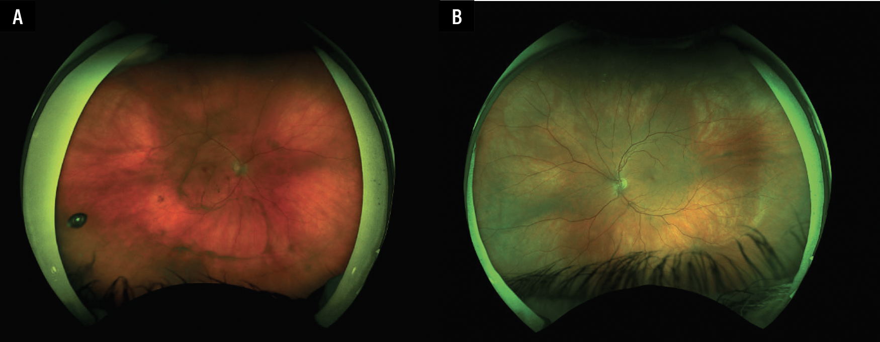 |
| Figure 1. Optos pseudocolor fundus photograph of the right eye (A) demonstrating presence of scattered retinal hemorrhages and vitritis. The left eye (B) has unremarkable findings. |
Initial workup and management
The patient was admitted to the hospital for a broad infectious and inflammatory workup, including chest X-ray, complete blood count, comprehensive metabolic panel, urinalysis, HIV serologies, syphilis IgG and rapid plasma reagin (RPR), QuantiFERON Gold, Toxoplasmosis IgG, and hepatitis C serologies, all of which were unremarkable. His bacterial and fungal blood cultures were additionally negative. He underwent vitreous tap and injection of voriconazole, ceftazidime and vancomycin, and the vitreous fluid was negative for bacterial and fungal culture and PCR, as well as viral (CMV, HSV and VZV) PCR. Gram stain of the vitreous fluid was notable for 3+ mononuclear cells. MRI orbits with and without contrast demonstrated unremarkable orbital findings. Our service obtained optical coherence tomography of the retina which didn’t demonstrate intraretinal or subretinal fluid in either eye.
Following discharge, he remained stable on topical steroids and atropine. Given the diagnostic uncertainty, he underwent diagnostic pars plana vitrectomy a month later. Flow cytometry, cytology, as well as bacterial and fungal culture and PCR were unremarkable. Pathology revealed scattered lymphocytes and histiocytes without findings consistent with malignancy.
Additional exam findings and workup completion
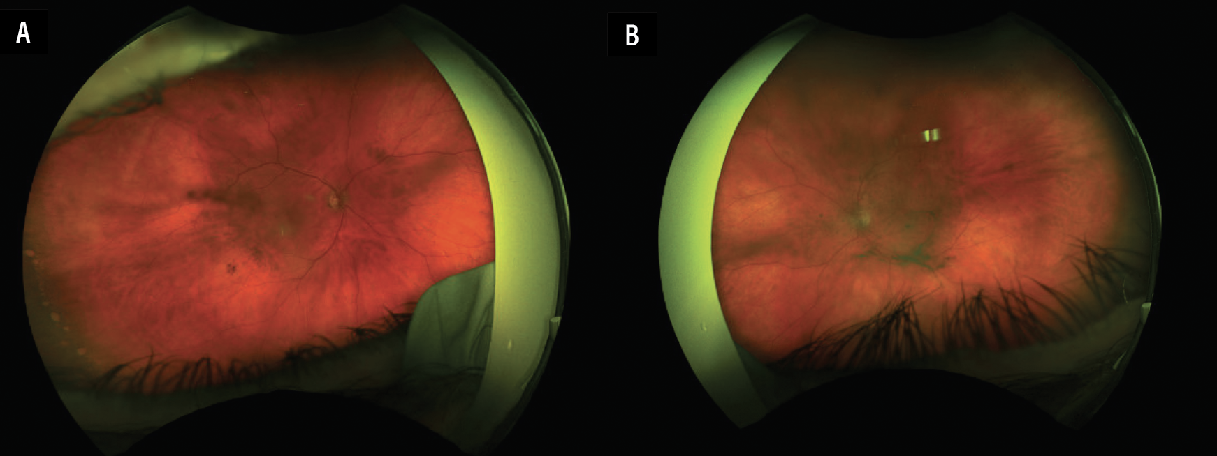
Two months following his initial PPV, he developed new floaters and blurred vision in the left eye. BCVA was 20/30 in the right eye and 20/50 in the left. Slit lamp and dilated fundus examination was unremarkable in the right eye, but revealed 1+ mixed anterior chamber cell, small keratic precipitates and 2+ clumped vitritis in the left eye (Figure 2). He subsequently underwent a diagnostic PPV of the left eye which similarly had unremarkable microbial studies, flow cytometry and cytology.
 |
|
Figure 2. Optos pseudocolor fundus photograph of the right eye demonstrates continued resolution of vitreous cell (A), while the photograph of the left eye shows new vitreous cell and haze partly obscuring details of the macula and optic nerve (B). |
In consultation with medical oncology, we obtained computed tomography scans of his chest, abdomen and pelvis, which were notable for multiple pulmonary nodules, retroperitoneal soft tissue nodules and radiolucent bone lesions. PET CT F-18 FDG whole body also demonstrated avid FDG uptake in the right supraclavicular lymph nodes and abdominal lymph nodes. Cytology from a retroperitoneal lymph node biopsy found monotonous round cells without specific immunologic differentiation markers, and flow cytometry was negative for abnormal B or T lymphoid cell populations, suggestive of dedifferentiated melanoma.
Diagnosis and management
He was diagnosed with bilateral paraneoplastic anterior and intermediate uveitis in the setting of stage IV metastatic dedifferentiated melanoma. His intraocular inflammation remained quiescent following his surgeries and he was started on palliative combination immunotherapy (anti-PD-1 plus anti-CTLA-4) for his systemic metastatic burden by oncology.
Discussion
Ocular paraneoplastic syndromes (OPS) are rare disorders that occur secondary to either immune-mediated cross-reactivity involving tumor antigens or ectopic production of hormones or growth factors by cancer cells.1 These syndromes may precede or accompany a recurrence in malignancy, highlighting their potential value as early warning signs. The estimated incidence of OPS in cancer patients is about 0.01 percent.2 While numerous malignancies are associated with OPS, the most common etiologies include small cell lung cancer, genitourinary malignancies such as endometrial and ovarian cancer, neuroendocrine tumors, lymphoma and malignant melanoma.3
Melanoma is associated with several systemic and ocular paraneoplastic syndromes. In a review of 59 cases, melanoma-associated retinopathy was the most common manifestation, while paraneoplastic encephalitis and cerebellar degeneration were less frequent.4 More rarely, melanoma-associated paraneoplastic vitelliform retinopathy has been described, characterized by serous macular detachment and nummular vitelliform retinal lesions.5
Paraneoplastic uveitis is a rare subtype of OPS. In a review of cases reported between 2010 and 2023, there were 11 cases of paraneoplastic uveitis associated with pulmonary malignancies and lymphoma identified, including three cases each of anterior, intermediate and posterior uveitis, and two cases of panuveitis.6 Other reports include bilateral hypopyon and vitritis as the initial manifestation of metastatic peritoneal carcinomatosis,7 and bilateral paraneoplastic vitritis without other fundus abnormalities in a patient with ocular small-cell lung carcinoma.8 An additional report described eight cases of granulomatous paraneoplastic vitritis, three of which led to a new cancer diagnosis and five of which signaled disease recurrence.9 Although the underlying malignancies were heterogeneous, none were associated with melanoma. This case represents one of the first reported cases of a paraneoplastic anterior and intermediate uveitis associated with malignant melanoma.
Metastatic melanoma portends a poor prognosis, with five-year survival rates estimated at about 35 percent for patients with distant metastases.10 Systemic treatments that can prolong life expectancy include checkpoint inhibitors such as anti-PD-1 and anti-CTLA-4 antibodies, which act to enhance the body’s T-cell ability to recognize and kill malignant cells. In our case, additional local therapy such as topical, intravitreal, or systemic steroids weren’t necessary in preventing recurrence of intraocular inflammation following PPV.
Bottom line
This case represents a unique presentation of metastatic melanoma presenting initially as an anterior and intermediate paraneoplastic uveitis. Clinicians should be aware of the malignancy-associated paraneoplastic syndromes, which facilitates earlier diagnosis and treatment. RS
REFERENCES
1. Przeździecka-Dołyk J, Brzecka A, Ejma M, et al. Ocular paraneoplastic syndromes. Biomedicines 2020;8:11:490.
2. Darnell RB, Posner JB. Paraneoplastic syndromes involving the nervous system. N Engl J Med 2003;349:16:1543-1554.
3. Alabduljalil T, Behbehani R. Paraneoplastic syndromes in neuro-ophthalmology. Curr Opin Ophthalmol 2007;18:6:463-469.
4. Dec M, Arasiewicz H. Paraneoplastic syndromes in patients with melanoma. Postepy Dermatol Alergol 2024;41:3:251-261.
5. Sotodeh M, Paridaens D, Keunen J, van Schooneveld M, Adamus G, Baarsma S. Paraneoplastic vitelliform retinopathy associated with cutaneous or uveal melanoma and metastases. Klin Monbl Augenheilkd 2005;222:11:910-914.
6. Beuzit S, Méal A, Delplanque M, et al. Paraneoplastic ocular syndromes: A systematic review of epidemiology, diagnosis and outcomes (2010-2023). J Ophthalmic Inflamm Infect 2025;15:1:73.
7. Iannetti L, Corsi C, Iafrate F, Sammartino P, Di Giorgio A, Pezzi PP. Bilateral uveitis with hypopyon as a presenting symptom of metastatic peritoneal carcinomatosis. Eur J Ophthalmol 2010;20:5:948-951.
8. Yigitaslan EK, Ucan ES, Akkoc N, Saatci AO. Bilateral paraneoplastic vitritis: A case report. Beyoglu Eye J 2021;6:2:140-144.
9. McGrath LA, Mudhar HS, Sheard R, et al. Paraneoplastic granulomatous vitritis: Elaboration of 8 Cases. Ophthalmol Retina 2019;3:7:589-596.
10. American Cancer Society. Survival rates for melanoma skin cancer. Updated January 13, 2026. Accessed May 19, 2026. https://www.cancer.org/cancer/types/melanoma-skin-cancer/detection-diagnosis-staging/survival-rates-for-melanoma-skin-cancer-by-stage.html.