 |
|
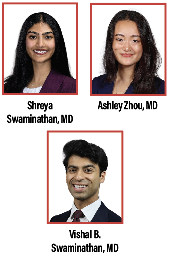
Bios Dr. Shreya Swaminathan is a PGY-2 ophthalmology resident at the Mayo Clinic in Rochester, Minn. |
The 14th Annual Vit-Buckle Society meeting brought retina specialists to Las Vegas with a “VBS in Wonderland” theme. This summary covers Session 9, which presented complex surgical cases and techniques, and Session 14, where surgeons debated controversial topics.
Session 9: Surgical Cases & Scientific Papers “Sometimes I’ve believed as many as six impossible things before breakfast.”
Moderators: Dean Eliott, MD & Gabriela Lopez Carasa, MD
Session 9 invited surgeons to present challenging cases and the techniques they used to manage them. The format highlights what worked, what didn’t, and what leaders in the field would do differently in difficult cases.
• Von Hippel-Lindau disease: capillary hemangioma–associated retinal detachment.
Lucas Zago, MD opened the session with a case of a 29-year-old woman with bilateral retinal capillary hemangiomas from Von Hippel-Lindau disease who presented with a tractional retinal detachment and hand motion vision. After treating the patient with preoperative anti-VEGF, Dr. Zago proceeded with a combined phacoemulsification and pars plana vitrectomy. He highlighted an iatrogenic break during the peeling process that prompted an extended superior retinectomy, as well as the need for endodiathermy to control an intraoperative bleed from the tumor. Vision reached 20/80 by month two and drifted to 20/200 by month nine.
Takeaway: Dr. Zago’s case emphasizes how residual hyaloid often hides beneath tractional membranes in VHL, and finding and addressing this may prevent re-detachment of the retina.
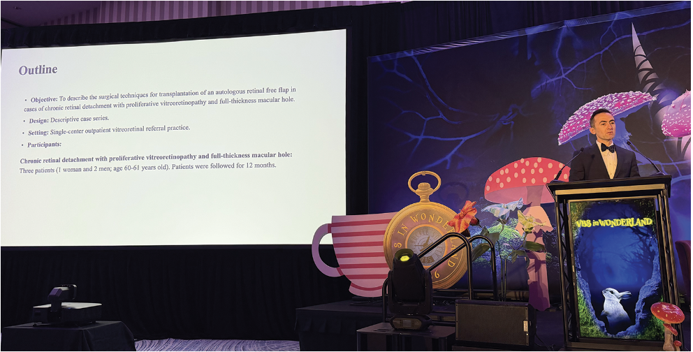 |
| Tarek Alasil, MD, discussed several cases pairing relaxing retinectomy with autologous retinal transplant. He found that in complex, macular hole-associated RD, tangential traction can undermine the success of the graft, underlining the need for the relaxing retinectomy. Photo: Matthew Starr, MD |
• What a mass! Exoresection for giant choroidal melanoma.
Yu-Bai Chou, MD, PhD described a globe-sparing transscleral resection technique in a 36-year-old woman with a giant (22.5 × 11.2 × 12.5 mm) choroidal melanoma and associated retinal detachment. With the patient electing to avoid enucleation, and radiotherapy not feasible at this size, Dr. Chou performed a posterior lamellar sclerouvectomy using a partial-thickness scleral flap and controlled suprachoroidal dissection for en bloc removal of the tumor, followed by a pars plana vitrectomy with silicone oil tamponade. At four years after resection, following oil removal and cataract surgery, the patient had 20/25 vision and, more importantly, globe preservation.
Takeaway: Dr. Chou showed the audience how globe-sparing exoresection is technically demanding but a viable alternative to enucleation in select patients with giant uveal melanomas. The audience voiced concerns regarding seeding of the orbit given exposure of the tumor cells to the orbital space during resection, but at this time, no recurrence had been noted.
• Into the rabbit hole: Macular hole repair in complex myopic detachment.
Aristomenis Thanos, MD presented a case of a highly myopic 62-year-old with a total macula-off retinal detachment with an inferonasal break and macular hole (VA 20/400). Dr. Thanos began the case with a combined phacoemulsification and pars plana vitrectomy, using a flex loop to help clear the adherent cortical vitreous. The posterior hyaloid was carefully addressed to release residual traction, and an amniotic membrane graft was positioned over the macular hole using preoriented markings. The retina remained stable after silicone oil removal, with graft removal planned.
Takeaway: In patients with high myopia and a macular hole with associated rhegmatogenous retinal detachment, amniotic membrane graft may be feasible to close the macular hole with concurrent RRD repair.
• Retinal redemption: Turning chaos into closure.
Bruna Gil Ferreira, MD, walked the audience through a series of chronic macula-off detachments with full-thickness macular holes and severe proliferative vitreoretinopathy. Dr. Ferreira described a case of a young patient with recurrent detachment where she began the repair with dissection of preretinal membranes using forceps and vertical scissors to release traction and mobilize the retina. After extensive PVR removal and a retinectomy, a free autologous retinal flap was created and positioned over the macular hole. Perfluorocarbon liquid stabilized the retina and guided flap placement before fluid-air exchange. Early imaging showed flap stabilization.
Takeaway: Dr. Ferreira highlighted how free autologous flaps are a great treatment method for treatment-refractory macular holes in advanced PVR, but functional recovery remains variable in published series.
• Surgical management for complex retinal detachment with PVR and macular hole using relaxing retinectomy and autologous retinal transplantation.
Tarek Alasil, MD, presented a series of complex detachment cases pairing relaxing retinectomy with autologous retinal transplantation. He presented a case of a patient who re-detached following a prior scleral buckle, in which extensive membrane peeling was conducted before he harvested an autologous graft and positioned it under perfluorocarbon over the macular hole with a flex loop. Endolaser was used to seal the donor site and a relaxing retinectomy released persistent traction. The patient had a good outcome with visual improvement. Additional cases showed modest but meaningful functional recovery.
Takeaway: In complex macular hole-associated retinal detachments, tangential traction can undermine the success of an autologous retinal graft, emphasizing the need for relaxing retinectomies.
• Macular hole management without tamponade.
Audrey Juan, MD, presented a gas-free approach to macular hole repair. After standard pars plana vitrectomy and ILM staining, she created a temporal ILM flap for large and extra-large holes. In place of gas, dispersive viscoelastic was layered over the flap at a low IOP to stabilize the flap. In her case series, six-month closure rates matched conventional gas tamponade, with faster early visual recovery in the no-gas group.
Takeaway: Viscoelastic over an ILM flap without gas may be a feasible option as more data is collected.
• First-in-human outcomes of a sutureless, scleral-fixated, prosthetic capsular bag.
Frank L. Brodie, MD, MBA, reported early outcomes with a sutureless scleral-fixated prosthetic capsular bag for secondary intraocular lens fixation. The device highlights the possibility of allowing a variety of IOLs to be chosen by the surgeon with flexible transscleral fixation arms that accommodate a range of axial lengths, in addition to integrated posterior vitrectomy ports. Across a 15-eye prospective series, there were no conjunctival erosions or IOL dislocations apart from a single footplate displacement in a patient with Marfan syndrome. Imaging showed stable centration with less tilt than Yamane fixation, approaching in-the-bag results.
Takeaway: A prosthetic bag may offer a more standardized solution for dislocated IOLs, with encouraging early data.
• Predictors of anatomic and functional success with amniotic membrane grafting for complex macular holes.
Scott D. Walter, MD, MSc, FASRS, presented a seven-year, single-surgeon retrospective case series on complex macular hole repair. He emphasized how there’s no universal approach to treatment and how intraoperative surgeon judgment should inform surgical technique. Use of silicone oil correlated with higher rates of complete and type 1 closure. Fibrin glue improved graft stability, particularly in complex or highly myopic eyes. Aggressive fluid-air exchange, by contrast, raised the risk of graft displacement.
Takeaway: For refractory holes, favor preretinal placement of amniotic membranes and use of silicone oil and fibrin glue, while avoiding aggressive fluid-air exchange that dislodges the graft.
• Outcomes of eyes with concurrent suprachoroidal hemorrhage and retinal detachment: A multicenter study.
Tianyu Liu, MD, presented a two-center retrospective study of eyes with concurrent suprachoroidal hemorrhage and retinal detachment from 2013 to 2022. At six months, visual outcomes were modest, with about 60 percent of eyes achieving reattachment overall. Rhegmatogenous cases typically required surgery for anatomic success, while some non-rhegmatogenous cases resolved without intervention. Proliferative vitreoretinopathy was consistently associated with worse anatomic and visual outcomes.
Takeaway: Dr. Liu emphasized in his talk how patients with PVR and rhegmatogenous retinal detachment may have a guarded prognosis in cases with concurrent suprachoroidal hemorrhage.
 |
Session 14: Surgical Retina Debates “I have an excellent idea! Let’s change the subject.” Moderators: Shilpa Desai, MD & Ella Leung, MD
The Saturday debates featured surgeons with opposing positions on clinically meaningful surgical questions, with the audience weighing in at the end of each debate.
• How to manage endophthalmitis: Tap-and-inject vs. immediate vitrectomy.
Samir N. Patel, MD, argued that tap-and-inject remains a practical first-line therapy in cases of endophthalmitis, citing modern data showing visual outcomes comparable to vitrectomy with lower procedural risk and broader accessibility. Maria H. Berrocal, MD, however, made the case for early vitrectomy, emphasizing rapid clearance of organisms, toxins and inflammatory mediators, with promising results due to early surgical intervention. Tap-and-inject narrowly took the room, 51 percent to 49 percent.
Takeaway: The Endophthalmitis Vitrectomy Study still guides practice for most cases, with a recent case series suggesting a lower threshold for early vitrectomy may result in better outcomes.
• Who can perform pediatric surgery: Specialty centers vs. all VR surgeons.
Nimesh A. Patel, MD, argued that pediatric vitreoretinal surgery belongs in dedicated children’s hospitals with the supporting infrastructure, such as pediatric anesthesia, child life services and coordinated perioperative care. Safa Rahmani, MD, on the other hand, countered that all VR surgeons should be prepared to handle at least some pediatric pathology, particularly trauma and retinal dialysis, given how limited specialty access can be for many families. The audience favored the points of Dr. Rahmani, agreeing there should be broader participation in pediatric retina surgery with a vote of 72 percent to 28 percent.
Takeaway: General VR surgeons should be comfortable managing pediatric trauma and retinal dialysis, however a referral to a dedicated pediatric hospital may be warranted in cases with complex pathology.
• How to manage RD: PPV alone vs. SB/PPV.
R. Ross Lakhanpal, MD, argued that small-gauge vitrectomy, wide-angle viewing, modern fluidics and improved illumination have reduced the need for scleral buckle procedures in primary retinal detachments, with faster recovery and fewer buckle-related complications. Talia R. Kaden, MD, made the case for the buckle in complex surgical scenarios, such as young phakic patients, cases with inferior pathology, and PVR or high-PVR-risk eyes. Dr. Kaden won, with 87 percent of the vote.
Takeaway: The Primary Retinal Detachment Outcomes Study data supports SB or combined PPV/SB over PPV alone in phakic eyes (91 to 92 percent vs. 83 percent single surgery anatomic success), especially with inferior breaks or PVR risk.
• Optimal timing for mac off RRD: The sooner, the better vs. there’s no urgency.
Ferhina S. Ali, MD, argued for earlier repair of macula-off retina detachments, citing meta-analyses showing improved outcomes with repair within three days of onset. Akshay S. Thomas, MD, MS, FASRS, pushed back and argued that repair should be timely, but most macula-off detachments don’t require overnight surgery and can be safely deferred several days. He emphasized how OR access, scheduling and patient expectations are all factors to consider. Dr. Thomas’ position in the debate carried the debate with a 87 percent vs. 13 percent.
Takeaway: Recent meta-analyses show better VA outcomes with repair within two to three days, but in the real world, pragmatic factors such as OR access, staffing and preoperative planning mean time and patience may allow for more optimal surgical preparation. RS