
 |
Bios
|
Post-traumatic dislocation of the crystalline lens into the subconjunctival space, referred to as phacocele, is a rare occurrence caused by an indirect scleral rupture of the globe. Cases of perforation due to ocular trauma are considered ophthalmic emergencies, requiring immediate and meticulous management.1 The presence of significant subconjunctival hemorrhage and hypotonia should raise suspicion for an occult perforation. B-scan ultrasonography is crucial for diagnosis, particularly in cases involving hyphema or vitreous hemorrhage. The absence of lens echoes on the scan is notable, and it also helps in identifying vitreous hemorrhages, retinal detachments, suprachoroidal hemorrhages and endophthalmitis.2
This report provides detailed insights into a case of phacocele and its outcome, while also addressing an important consideration in this condition, which is primary versus secondary intraocular lens implantation.
Surgical management
This patient presented to our clinic 10 days after being struck in the right eye by an iron bar, reporting pain and vision loss (limited to light perception). A slit-lamp exam revealed a solid, globular yellowish mass in the subconjunctival space in the superonasal quadrant, with a smooth surface and rounded margins. Visualization of the anterior chamber was obstructed by corneal edema and a 3.5-mm hyphema. Aphakia and iris atrophy from 10 to 1 o’clock were noted, with an intraocular pressure of 17 mmHg and no relative afferent pupillary defect. B-scan ultrasonography showed dispersed hemorrhage in the anterior vitreous and the absence of a lens spike, confirming the diagnosis of traumatic phacocele.
Explorative surgery was performed under retrobulbar anesthesia. A 360° conjunctival peritomy was conducted, and the lens with its intact capsule was removed from the subconjunctival space. A 7-mm superior scleral wound with uveal tissue remnants was observed. Although a human scleral graft was prepared, it wasn’t needed due to the regular and linear nature of the wound. A standard three-port pars plana vitrectomy was then performed, followed by scleral introflexion to alleviate potential residual tractions at the wound site. During the vitrectomy, multiple areas of scar fibrosis with hyperpigmented, well-defined grayish lesions were identified along the retina. The eye was left aphakic to allow for better surgical preparation in a subsequent procedure. At the one-month follow-up, the best-corrected visual acuity of the right eye was 20/60.
Case takeaways
The most frequent location for a phacocele is the superonasal quadrant near the limbus, attributed to the energy projection from an impact in the temporal region. The superotemporal area is the second most common site. Scleral ruptures typically occur between the limbus and the spiral of Tillaux.3,4 Phacocele is more commonly observed in elderly individuals with a rigid sclera compared to children, who have more elastic ocular tissues. These cases are often accompanied by vitreous hemorrhage and retinal detachments.5,6
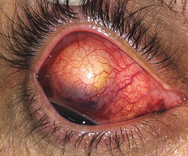 |
| Figure 1. External photography of the right eye, highlighting the boundaries of the subconjunctival mass. |
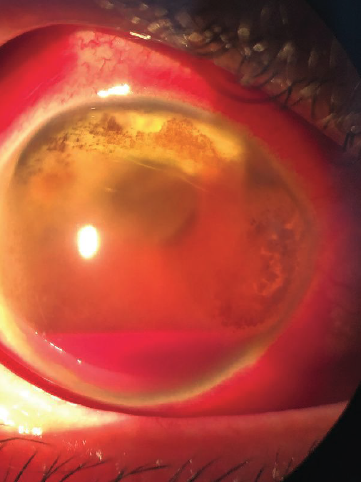 |
| Figure 2. Slit-lamp examination showing corneal edema, hyphema of 3.5 mm height, aphakia and iris atrophy from 10 o’clock to 1 o’clock. |
The main objective in managing a phacocele is to remove the displaced lens while preserving the integrity of the globe.7 In surgical management, it’s recommended to first place the infusion port to prevent eye collapse during manipulation of the lens near the scleral wound, followed by exploration of the injury. The scleral rupture should be repaired, and partial vitrectomy performed if needed. Scleral introflexion is advised to alleviate any residual traction, given the complexity of treating the vitreous base in these cases. IOL implantation can be deferred to a later session based on the condition of the eye.
One paper found that primary IOL implantation, using biometry from the non-traumatized contralateral eye to calculate IOL power, could lead to a significant refractive error of up to 4 D. However, after revising the approach and performing IOL exchange surgery using simulated keratometry and axial length measurements from the injured eye, the patient achieved emmetropia with a BCVA of 20/20.8 On the other hand, another report emphasized that using the biometry of the injured eye after primary repair of the globe was more accurate than using biometry of the non-traumatic eye to determine the IOL power with refraction deviation of only 1 D in secondary IOL implantation.9 Further, timely and effective intervention can ensure good visual recovery.
In the case reported here, the primary objective of the surgery was to preserve the integrity of the globe, allowing for a future procedure with optimized IOL power calculation under the best possible conditions. RS
REFERENCES
1. Can HY, Koru M, Karahan E. A traumatic phacocele after blunt trauma: A case report. J Med Cases Updat 2021;1:1:10-11. http://jmcupdates.com/index.php/jmc/article/view/7. Accessed December 22, 2021.
2. Neupane S, Das D, Korgaonkar S. Migrated subconjunctival crystalline lens: A traumatic phacocele. BMJ Case Rep 2020;13:7.
3. Allen RC, Gupta RR, Poblete R, Oetting TA. Traumatic phacocele. J Cataract Refract Surg 2001;27:8:1333-1334.
4. Sindal MD, Mourya D. A rare case of traumatic posterior phacocele with retinal detachment. Indian J Ophthalmol 2016;64:1:89-90.
5. Bhattacharjee K, Bhattacharjee H, Deka A, Bhattacharyya P. Traumatic phacocele: Review of eight cases. Indian J Ophthalmol 2007;55:6:466-468.
6. Santos-Bueso E, Sáenz-Francés F, Díaz-Valle D, et al. [Ocular rupture associated with lens dislocation to the subconjunctival space]. Arch Soc Esp Oftalmol 2007;82:10:641-644.
7. Sathish S, Chakrabarti A, Prajna V. Traumatic subconjunctival dislocation of the crystalline lens and its surgical management. Ophthalmic Surg Lasers 1999;30:8:684-686.
8. Cohen KL. Inaccuracy of intraocular lens power calculation after traumatic corneal laceration and cataract. J Cataract Refract Surg 2001;27:9:1519-1522.
9. Lan-Hsin C, Chi-Chun L. Secondary intraocular lens implantation of traumatic cataract in open-globe injury. Can J Ophthalmol 2005;40:4:454-459.