Take-home points
|
 |
Bio Dr. Yokoi is a former postdoctoral research fellow at Wills Eye Hospital and is currently chief doctor in the Department of Ophthalmology at the National Center for Child Health and Development in Tokyo. |
Managing pediatric retinal detachment requires a disease- and anatomy-specific strategy rather than a scaled-down application of adult retinal detachment surgery. The pediatric eye has a developing pars plana, a proportionally large crystalline lens, a formed and often firmly adherent vitreous, and a higher likelihood of developmental or inherited vitreoretinal disease.1,2 In rhegmatogenous retinal detachment, particularly in phakic children with anterior pathology, scleral buckling should be considered as a primary procedure.3-5 In tractional retinal detachment, the priority is controlled release of clinically significant traction while avoiding maneuvers that may create iatrogenic breaks,1,2,5 an approach that embodies the “Less is more” philosophy.
Examination under anesthesia and setting up the case
Examination under anesthesia is often part of the operation rather than a separate diagnostic step. The surgeon should document the retina with scleral depression, examine the fellow eye, obtain OCT or ultrasonography when useful, and perform widefield fluorescein angiography when FEVR, ROP or peripheral ischemia is suspected. Before placing a trocar for vitrectomy, the entry site should be selected for that child. Craig A. Lemley, MD, and Dennis P. Han, MD, proposed 1.5 mm posterior to the limbus at 1 to 6 months, 2 mm at 6 to 12 months, 2.5 mm at 1 to 2 years, 3 mm at 2 to 6 years, and 3.5 mm at 6 to 18 years. Lauren Wright, MD, and colleagues later emphasized that disease-specific anatomy, including ROP, PFV, coloboma and high myopia, can modify these distances.6,7
Infant trocar placement shouldn’t be performed like routine adult transconjunctival entry. After conjunctival cutdown, the intended sclerotomy site should be stabilized by grasping the adjacent sclera with forceps, allowing controlled trocar penetration despite the extremely soft sclera. A small pre-incision with a sharp blade, such as a V-lance, may further improve control in selected cases. The trocar should be directed more vertically, while carefully avoiding the lens. At the end of surgery, ports should be sutured liberally in neonates, infants, young children, hypotonous eyes or children likely to rub the eye postoperatively.
Visualization is a working-space issue as much as an optical issue. In infants, narrow fissures and small globes with shorter pars plana may cause a standard round noncontact lens to interfere with instrument movement. Smaller-footprint Resight-compatible front lenses can be useful, including the Oculus Versa HD Disposable LenZ for Zeiss Resight and Picture Technology’s PT-S133 in Japan.
Shorter instruments can be considered, especially for the neonate and infant cases. Commercially available short-shaft systems, such as the Constellation 25+ Short Ultravit Vitrectomy Probe with compatible short-pak instrumentation (Alcon) or ultra-short vitrectomy kits from DORC, may be useful, particularly in neonatal and infant eyes. These instruments, with an approximately 18-mm shaft, can improve maneuverability and reduce the risk of excessive intraocular entry.
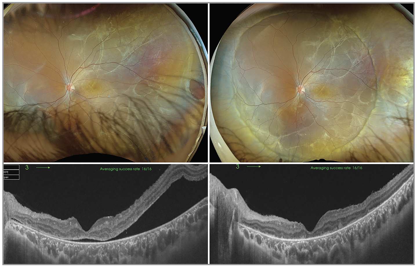 |
| Figure 1. An 11-year-old girl with subretinal PVR in the left eye caused by a peripheral retinal break at 3 o’clock associated with FEVR. The left panels show the preoperative pseudocolor fundus photograph and OCT image. BCVA was 20/200, and both the fundus photograph and OCT findings indicated that the detachment was longstanding. Encircling scleral buckling was performed using a #506 sponge. The right panels show successful retinal reattachment and improvement of BCVA to 20/60 at one month postop. This case illustrates the importance of scleral buckling, even in a complicated case with subretinal PVR. |
RRD: Consider buckling first
Many pediatric RRDs begin anteriorly: dialysis; ora breaks; lattice holes; traumatic tears; Stickler-related vitreous-base abnormalities; or FEVR-associated peripheral breaks. In these eyes, scleral buckling should be actively considered before primary vitrectomy, even in the presence of subretinal PVR, as illustrated by the representative case shown in Figure 1. Pediatric RRD series support primary scleral buckling, and primary vitrectomy alone may be less favorable than scleral buckle or combined buckle-vitrectomy in selected children.3-5 A segmental buckle is appropriate for the RD due to a localized lattice hole(s). Because many pediatric vitreoretinal diseases involve broad circumferential peripheral pathology, an encircling buckle should be considered for multiple holes, inferior pathology, high myopia, Stickler syndrome without a giant retinal tear, FEVR or trauma.
Vitrectomy is often unavoidable for giant retinal tear, posterior break, dense vitreous hemorrhage, severe PVR, especially with severe preretinal proliferation; traumatic incarceration, breaks with highly liquefied vitreous or failed buckle.3,8 Although complete PVD induction may be desirable, it shouldn’t be forced when anomalous, firm vitreoretinal adhesion makes the posterior hyaloid inseparable. Careful shaving, traction segmentation and external support may be safer than aggressive posterior hyaloid separation.1,2 In pediatric giant retinal tear, stabilize the posterior flap with perfluorocarbon liquid, meticulously trim anterior flap traction, and strongly consider encircling buckling in young children, syndromic eyes or eyes with inferior pathology.8 Gas may be appropriate for an older cooperative child with superior breaks. Silicone oil is often more practical in young children, in eyes with inferior pathology, PVR, giant retinal tears, or combined TRD/RRD, and when reliable postoperative positioning is unlikely.
In Stickler syndrome, Cambridge cryotherapy and Manchester 360-degree laser prophylaxis have both reported substantial reduction in detachment risk, and established detachments may require multiple procedures with combinations of buckle, vitrectomy, retinopexy and silicone oil.9-11 During vitrectomy, a thin residual posterior hyaloid may persist despite extensive vitreous liquefaction. Its identification and removal are often critical to success. Careful shaving of the thick peripheral vitreous over areas of characteristic radial perivascular degeneration is also important.
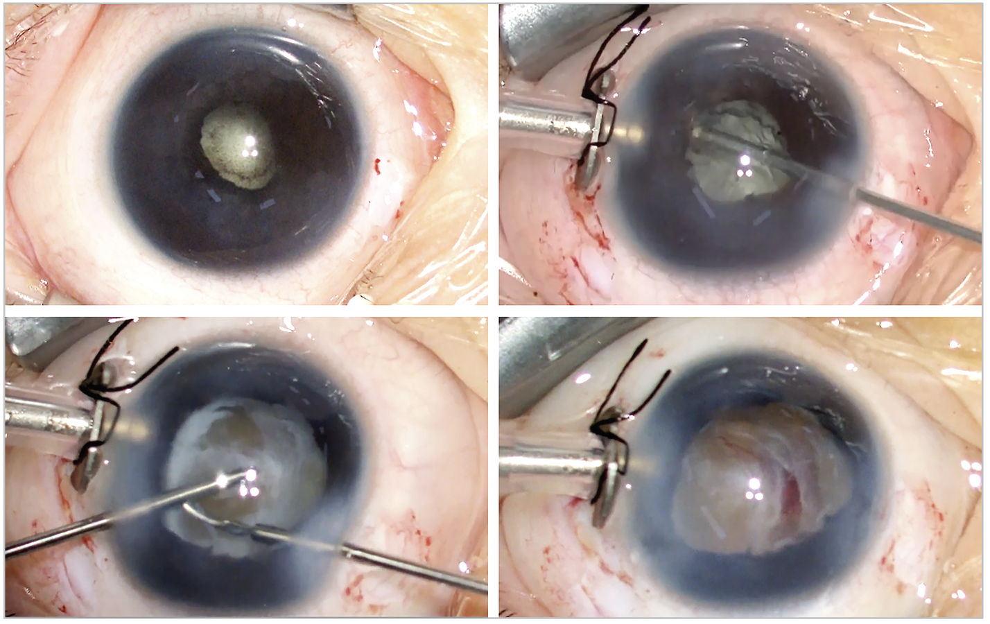 |
| Figure 2. An 11-month-old boy with stage 5 FEVR in the left eye, shown from the surgeon’s view. The corneal diameter was 9 mm, slightly smaller than normal. The iris was partially adherent to the cornea superiorly, laterally, and inferiorly, with mild corneal opacity in these areas. Preoperatively, a retrolental fibrovascular plaque pushed the lens anteriorly, with 360-degree posterior synechiae and an extremely shallow anterior chamber (top left). A specially designed 2.5-mm infusion cannula was placed through the limbus and sutured, and lensectomy was performed after dissection of the iridocorneal adhesions and sphincterectomy (top right). The retrolental fibrous tissue was dissected and resected (bottom left), and the completely closed funnel was opened toward the optic nerve head. A certain amount of preretinal fibrous membrane was intentionally left in place to avoid iatrogenic breaks, consistent with the “less is more” philosophy. |
FEVR can produce RRD, TRD, or a combined detachment. Avascular peripheral retina and anomalous vascular development should be assessed with widefield imaging.12 If peripheral holes and anterior contraction dominate, encircling buckling should be considered first, together with treatment of ischemic retina when indicated13 When traction around the causative break is substantial, combined scleral buckling and vitrectomy is often required. In FEVR-associated RRD, prognosis depends largely on the break location and the surrounding traction. Breaks located beneath or adjacent to retinal folds make surgical repair considerably more complex and the visual prognosis more guarded.
RD in morning glory disc anomaly and chorioretinal coloboma often require vitrectomy, because the pathologic communication and causative break may lie at the disc anomaly, intercalary membrane or coloboma margin. Around anomalous optic discs, the posterior hyaloid is often abnormal and tightly adherent. Complete PVD induction is frequently impossible; therefore, anomalous vitreous should be removed conservatively rather than by forceful hyaloid separation.14-16 In coloboma-associated RD, search carefully for breaks in the intercalary membrane and at the coloboma margin, apply laser to identified breaks if possible, and the coloboma edge, and use long-acting tamponade in unstable eyes.15,16 In morning glory anomaly, peripapillary communication and abnormal disc tissue can make reattachment slow and recurrence possible; surgery often requires careful vitrectomy, cautious peripapillary laser, and silicone oil in highly complex cases.14 Because the use of silicone oil in eyes with chorioretinal coloboma or morning glory anomaly remains controversial owing to the potential risk of intracranial migration, explicit preoperative discussion with the parents is essential.
TRD: Selective release rather than exhaustive peeling
Pediatric TRD surgery is controlled traction release. In ROP, FEVR and PFV, the goal isn’t to remove every membrane but to reduce the traction that threatens or distorts the macula. Firmly adherent tissue may be left in place when peeling would create a retinal break. Once an iatrogenic break is created, visual outcomes become less favorable; therefore, each maneuver should be judged by whether it safely improves the retinal configuration and visual potential.5
 |
In ROP, stage 4A is the most favorable surgical window: The macula is still attached, and lens-sparing vitrectomy can relax ridge traction before the eye progresses to a funnel configuration.17,18 PVD induction isn't required. Traction should be segmented along ridge-to-ora, ridge-to-lens and circumferential vectors. Because laser photocoagulation of the far peripheral retina can be difficult during lens-sparing vitrectomy, preoperative external laser treatment should be considered when indicated. If anti-VEGF has been used, vascular activity may decrease while fibrotic contraction continues or even accelerates; timing should be guided by the tractional course and retinal configuration rather than the injection date alone.17
Stage 5 ROP and FEVR shift the surgical access problem. When anterior traction, retrolental fibroplasia, or a closed funnel prevents safe pars plicata work, an anterior lensectomy-vitrectomy approach, or limbal approach, may provide a far safer surgical corridor than pars plicata trocar placement.19 The aim is to open the funnel and free the posterior pole when possible. A partially relaxed posterior pole without iatrogenic breaks may be a better endpoint (Figure 2) than an extensive dissection that converts TRD into combined TRD/RRD.
FEVR-TRD is age dependent. Younger children may have more vascularly active proliferation, whereas older children often have more fibrotic folds. Ischemic retina with neovascular components should be treated with laser when accessible, buckle support should be considered for peripheral contraction, and vitrectomy should be used for posterior tractional maculopathy. The appropriate endpoint is reduction of macular distortion, not complete removal of every peripheral membrane.20 Most importantly, adequate laser photocoagulation of ischemic retina is fundamental to controlling vascular activity and achieving long-term success in this disease.
PFV is caused by abnormal persistence of the fetal hyaloid vascular system. The retrolental plaque, stalk, ciliary processes, anterior retina and posterior pole may be connected. Early surgery can help selected posterior PFV eyes with tractional detachment and tractional maculopathy, but microphthalmia, glaucoma, cataract and macular aplasia or dysplasia limit prognosis.21-23 If anterior displacement of the retina or dense retrolental tissue obscures the route, limbal access may be safer than pars plana entry.22 Lens preservation is valuable only when it still permits meaningful release of stalk and macular traction. For isolated anterior PFV, early surgery is often favored by analogy with the timing principles of unilateral congenital cataract surgery. In contrast, vitrectomy for posterior or combined anterior-posterior PFV is indicated more selectively, depending primarily on macular involvement.
In conclusion, successful pediatric retinal detachment surgery begins with understanding the pediatric vitreous, the developing retina and the disease-specific mechanisms that shape each detachment. RS
REFERENCES
1. Gan NY, Lam WC. Special considerations for pediatric vitreoretinal surgery. Taiwan J Ophthalmol 2018;8:4:237–242.
2. Yonekawa Y, Fine HF. Practical pearls in pediatric vitreoretinal surgery. Ophthalmic Surg Lasers Imaging Retina 2018;49:8:561–565.
3. Smith JM, Ward LT, Townsend JH, et al. Rhegmatogenous retinal detachment in children: Clinical factors predictive of successful surgical repair. Ophthalmology 2019;126:9:1263–1270.
4. Errera MH, Liyanage SE, Moya R, Wong SC, Ezra E. Primary scleral buckling for pediatric rhegmatogenous retinal detachment. Retina 2015;35:7:1441–1449.
5. Read SP, Aziz HA, Kuriyan A, et al. Retinal detachment surgery in a pediatric population: Visual and anatomic outcomes. Retina 2018;38:7:1393–1402.
6. Lemley CA, Han DP. An age-based method for planning sclerotomy placement during pediatric vitrectomy: A 12-year experience. Trans Am Ophthalmol Soc 2007;105:86–89. Discussion 89–91.
7. Wright LM, Harper CA, Chang EY. Management of infantile and childhood retinopathies: Optimized pediatric pars plana vitrectomy sclerotomy nomogram. Ophthalmol Retina 2018;2:12:1227–1234.
8. Ong SS, Ahmed I, Gonzales A, et al. Vitrectomy versus vitrectomy with scleral buckling in the treatment of giant retinal tear related retinal detachments: An international multicenter study. Ophthalmol Retina 2022;6:7:595–606.
9. Fincham GS, Pasea L, Carroll C, et al. Prevention of retinal detachment in Stickler syndrome: The Cambridge prophylactic cryotherapy protocol. Ophthalmology 2014;121:8:1588–1597.
10. Linton E, Jalil A, Sergouniotis P, et al. Laser prophylaxis in Stickler syndrome: The Manchester protocol. Retina 2023;43:1:88–93.
11. Abeysiri P, Bunce C, da Cruz L. Outcomes of surgery for retinal detachment in patients with Stickler syndrome: A comparison of two sequential 20-year cohorts. Graefes Arch Clin Exp Ophthalmol 2007;245:11:1633–1638.
12. Tauqeer Z, Yonekawa Y. Familial exudative vitreoretinopathy: Pathophysiology, diagnosis, and management. Asia Pac J Ophthalmol (Phila) 2018;7:3:176–182.
13. Huang L, Liang T, Lyu J, Jin H, Zhao P. Clinical features and surgical outcomes of encircling scleral buckling with cryotherapy in familial exudative vitreoretinopathy-associated rhegmatogenous retinal detachment. Retina 2022;42:1:55–63.
14. Chang S, Gregory-Roberts E, Chen R. Retinal detachment associated with optic disc colobomas and morning glory syndrome. Eye (Lond) 2012;26:4:494–500.
15. Gopal L, Badrinath SS, Sharma T, et al. Surgical management of retinal detachments related to coloboma of the choroid. Ophthalmology 1998;105:5:804–809.
16. Abouammoh MA, Alsulaiman SM, Gupta VS, et al. Surgical outcomes and complications of rhegmatogenous retinal detachment in eyes with chorioretinal coloboma: The results of the KKESH International Collaborative Retina Study Group. Retina 2017;37:10:1942–1947.
17. Kusaka S. Current concepts and techniques of vitrectomy for retinopathy of prematurity. Taiwan J Ophthalmol 2018;8:4:216–221.
18. Lakhanpal RR, Sun RL, Albini TA, Holz ER. Anatomic success rate after 3-port lens-sparing vitrectomy in stage 4A or 4B retinopathy of prematurity. Ophthalmology 2005;112:9:1569–1573.
19. Sen P, Jain S, Bhende P. Stage 5 retinopathy of prematurity: An update. Taiwan J Ophthalmol 2018;8:4:205–215.
20. Yamane T, Yokoi T, Nakayama Y, Nishina S, Azuma N. Surgical outcomes of progressive tractional retinal detachment associated with familial exudative vitreoretinopathy. Am J Ophthalmol 2014;158:5:1049–1055.
21. Bosjolie A, Ferrone P. Visual outcome in early vitrectomy for posterior persistent fetal vasculature associated with traction retinal detachment. Retina 2015;35:3:570–576.
22. Ozdek S, Ozdemir Zeydanli E, Atalay HT, Aktas Z. Anterior elongation of the retina in persistent fetal vasculature: Emphasis on retinal complications. Eye (Lond) 2019;33:6:938–947.
23. Prakhunhungsit S, Berrocal AM. Diagnostic and management strategies in patients with persistent fetal vasculature: Current insights. Clin Ophthalmol 2020;14:4325–4335.
24. Hirakata A, Inoue M, Hiraoka T, McCuen BW. Vitrectomy without laser treatment or gas tamponade for macular detachment associated with an optic disc pit. Ophthalmology 2012;119:4:810–818.