Take-home points
|
 |
Bios Dr. Garg is chief of the Retina Service at Wills Eye Hospital. He practices at Mid Atlantic Retina and is a professor of ophthalmology at Sidney Kimmel Medical College at Thomas Jefferson University. |
The treatment landscape for geographic atrophy secondary to age-related macular degeneration has undergone a dramatic transformation with the U.S. Food and Drug Administration approval of complement-inhibiting therapies. Despite these advances, significant unmet needs remain, as current treatments modestly slow lesion growth but, unfortunately, they can’t restore vision. A broad range of therapeutic strategies are under active investigation, including complement pathway modulation, visual cycle inhibition, antioxidative therapies, reduction of toxic cellular byproducts, neuroprotection, laser-based interventions, photobiomodulation, surgical implants, gene therapy, stem cell therapies and mitochondrial enhancement. In this article, we’ll discuss some of the new treatment programs currently in development.
Complement inhibition: The current standard and future directions
Dysregulation of the complement cascade remains one of the most extensively studied mechanisms in GA pathogenesis. The two currently approved therapies target different components of the complement system: pegcetacoplan targets C3 and C3b and avacincaptad pegol targets C5.
•Pegcetacoplan. Pegcetacoplan is a pegylated peptide that binds complement C3/C3b and prevents downstream complement activation. In the Phase III OAKS trial, at 12 months, monthly and every-other-month intravitreal administration reduced overall GA lesion growth (foveal and non-subfoveal lesions combined) by 21 percent and 16 percent, respectively. Although the companion DERBY trial failed to meet its primary endpoint at 12 months, longer-term follow-up demonstrated benefit in both studies. At 24 months, monthly treatment reduced lesion growth by 22 percent in OAKS and 19 percent in DERBY, while every-other-month dosing reduced growth by 18 percent and 16 percent, respectively. This drug has also been approved by the FDA.
The ongoing GALE extension study has shown increasing treatment benefit over time, with up to a 42-percent reduction in lesion growth versus projected sham in the monthly treatment arm compared to projected sham at 36 month.1
The GARLAND study (Apellis) is a Phase IV real-world study evaluating pegcetacoplan's long-term efficacy and safety. This is a post-marketing observational study that's expected to be completed in June 2027.
•Avacincaptad pegol. The Phase II/III GATHER1 study demonstrated that monthly intravitreal avacincaptad pegol reduced non-subfoveal GA lesion growth by 27 percent (35 percent when square-root transformation wasn’t used) at 12 months compared with sham treatment.2 These findings were subsequently supported by the Phase III GATHER2 trial, which reported a 14-percent reduction (18 percent if not calculated using square-root transformation) in non-subfoveal GA area growth at 12 months with monthly dosing.3 Two-year data was recently published and supported efficacy for both monthly (14-percent difference versus sham) and every-other-month (19-percent difference versus sham) dosing.4 These studies established C5 inhibition as an effective strategy for slowing GA progression and led to US FDA approval.
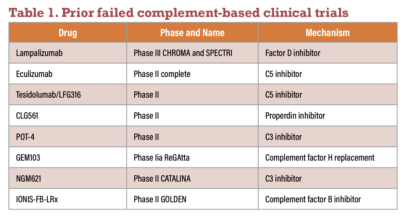 |
Ongoing complement-based trials
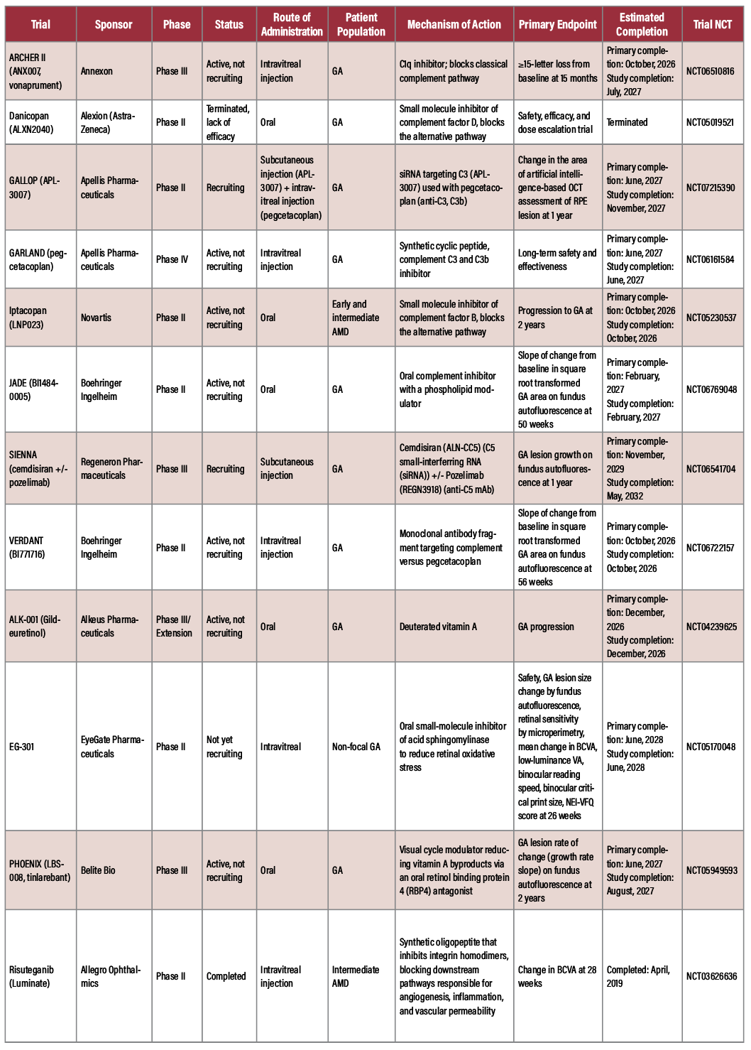
Several other complement inhibitors are in clinical development. We highlight a few of them here. There are several others in the pipeline, so please see clinicaltrials.gov for a more comprehensive evaluation.
 |
• Phase III programs. The Archer II (Annexon) is a study of an intravitreal complement inhibitor called vonaprument that’s being evaluated for patients with central GA. Unlike the current treatments, it targets C1q, an essential part of the classic pathway but leaves the alternative and lectin pathways alone hopefully allowing normal immune function to occur more fully.
The Phase II ARCHER study failed to reduce GA lesion growth at 12 months but demonstrated significantly less visual function loss. Only 6 percent of treated eyes lost ≥15 letters compared with 21 percent of sham eyes. The ongoing Phase III ARCHER II trial is evaluating ≥15-letter vision loss at 18 months as its primary endpoint.
The SIENNA trial (Regeneron) is a Phase III study evaluating systemic complement inhibition for patients with GA. The study uses cemdisiran, a small interfering RNA (siRNA) that suppresses complement component C5, with or without pozelimab, a monoclonal antibody against C5. By inhibiting C5, the treatment aims to prevent formation of the membrane attack complex and reduce complement-mediated retinal damage.
The primary endpoint is GA lesion growth measured by fundus autofluorescence at one year. Unlike currently approved intravitreal complement inhibitors, SIENNA is investigating a subcutaneous systemic therapy, which may reduce treatment burden while providing sustained complement inhibition.
• Phase II programs. The GALLOP trial (Apellis Pharmaceuticals) is a Phase II study evaluating a combination complement inhibition strategy for patients with GA. Participants receive APL-3007, a subcutaneous siRNA that suppresses complement component C3 production, together with intravitreal pegcetacoplan, an approved C3/C3b inhibitor. The goal is to determine whether dual systemic and local inhibition provides greater protection against retinal degeneration than intravitreal therapy alone.
The primary endpoint is the change in retinal pigment epithelium lesion area measured by artificial intelligence-based OCT analysis at one year. This study is among the first to evaluate whether combining systemic and intravitreal complement inhibition can improve treatment outcomes in GA.
The VERDANT trial (Boehringer Ingelheim) is a Phase II study evaluating BI771716, an intravitreal monoclonal antibody fragment targeting the complement pathway, in patients with GA. The study directly compares BI771716 with pegcetacoplan, making it one of the few head-to-head trials between investigational and approved complement therapies.
The primary endpoint is the slope of change in square root-transformed GA lesion area on fundus autofluorescence through 56 weeks. The trial is designed to determine whether BI771716 can slow GA progression as effectively as, or better than, the current standard of care while further characterizing its safety profile.
The JADE study (Boehringer Ingelheim) is a Phase II study evaluating BI1484-0005, a daily oral PAF receptor antagonist. BI1484-0005 targets the PAF receptor which is involved in inflammation, angiogenesis and vascular permeability. The primary endpoint is slope of change from baseline in square-root transformed GA area, as measured by FAF at 50 weeks.
Another Phase II trial is evaluating iptacopan (LNP023), an oral complement factor B, for patients with early and intermediate AMD. This trial is evaluating the progression to GA at two years.
 |
| Click image to enlarge. |
 |
| Click image to enlarge. |
Visual cycle modulation
Visual cycle modulators aim to reduce accumulation of toxic vitamin A-derived byproducts implicated in retinal pigment epithelium dysfunction and photoreceptor loss.
ALK-001 (gildeuretinol), a deuterated vitamin A derivative, is designed to reduce vitamin A dimerization and subsequent formation of toxic retinal byproducts. Although the therapy didn’t demonstrate a statistically significant reduction in GA growth through 24 months, post hoc analyses suggested a 15.3-percent reduction in lesion growth between months six and 24. Further studies may clarify whether earlier intervention or patient selection could enhance treatment benefit. The Phase III study is fully enrolled, and results are expected in December 2026.
Belite Bio’s Phase III PHOENIX study also targets the visual cycle. This oral therapy aims to reduce the accumulation of toxic vitamin A-derived byproducts implicated in retinal degeneration and may offer a noninvasive treatment alternative for patients with GA. The PHOENIX study is fully enrolled, and results are expected in August 2027.
Antioxidative therapies
Oxidative stress remains a central mechanism in AMD pathogenesis. Antioxidant supplementation has long been investigated as a strategy to slow disease progression.
The landmark AREDS trial demonstrated a 25-percent reduction in progression to advanced AMD with antioxidant and zinc supplementation. However, this benefit was primarily driven by reduced progression to neovascular AMD rather than prevention of GA enlargement. AREDS2 subsequently replaced β-carotene with lutein and zeaxanthin and suggested a potential slowing of central GA progression.5 In a post-hoc analysis of AREDS2, patients with non-central GA experienced slower rate of growth toward the center of the macula.6
Current investigations include EG-301 (EG-DPMP-01, Evergreen Therapeutics, NCT05170048), which is undergoing Phase II evaluation to slow GA area expansion at 26 weeks. This is an oral therapy that targets choriocapillaris, retinal and RPE oxidative stress, though the exact mechanism remains proprietary.
Reduction of toxic cellular byproducts
Accumulation of toxic protein aggregates and retinal waste products may contribute to RPE degeneration and photoreceptor death. GAL-101 (MRZ-99030, Galimedix Therapeutics, NCT06659549) is being investigated via the eDREAM study. This is a topical ophthalmic drop that targets beta amyloid oligomers. Primary completion is expected in March 2027.
This class of therapy seeks to reduce cellular toxicity and preserve retinal structure before irreversible atrophy develops.
Neuroprotective therapies
Neuroprotection represents another promising strategy aimed at preserving photoreceptors and retinal neurons despite ongoing disease processes.
Risuteganib (Luminate, ALG-1001) is currently being evaluated in a Phase II clinical trial. By targeting integrin signaling pathways, risuteganib may improve cellular resilience and retinal function independent of complement modulation. Risuteganib is thought to function by binding to RPE cells to reduce mitochondrial reactive oxygen species, reducing RPE cell death due to oxidative stress.7
Laser-based therapies
Laser interventions are being explored as potential methods to modulate retinal metabolism and stimulate protective cellular responses. The Laser Intervention in Early Stages of Age-Related Macular Degeneration (LEAD) study was a 36-month trial conducted in Australia that evaluated subthreshold nanolaser in 292 patients with intermediate AMD to assess the progression to GA. Subthreshold nanolaser produces a non-visible laser spot that’s thought to function by modulating RPE-mediate turnover of, and reduce outflow resistance at, Bruch’s membrane. This study failed to meet its primary endpoint, though post-hoc analysis suggests patients without reticular pseudodrusen may experience slowed progression to GA.8 Another clinical trial is underway in Australia evaluating the R:GEN Laser System (LASER LIGHT-01) on progression to GA.9
Photobiomodulation
Photobiomodulation uses specific wavelengths of light to improve mitochondrial function and reduce oxidative stress.
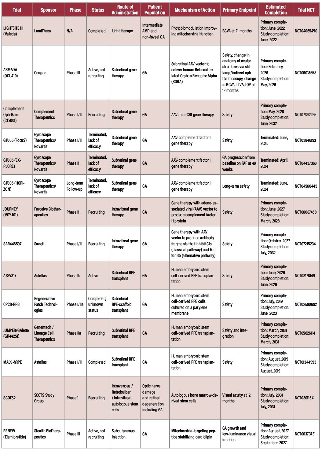
The Valeda Light Delivery System has been evaluated in the LIGHTSITE I, II and III clinical trials. The LIGHTSITE III study enrolled 148 eyes with intermediate dry AMD or non-foveal GA which and randomized them to receive either photobiomodulation or sham therapy, three times per week over three to five weeks, every four months for two years (total of 52 treatment sessions). Study patients exhibited significantly lower incidence of progression compared to sham (6.8 versus 24 percent).10 The proposed mechanism for PBM is mitochondrial photoreceptor activation to produce a biologic response that stabilizes metabolic function, promotes cytoprotection and produces energy.10
Surgical and implantable technologies
Advanced visual prosthetic and implantable technologies seek to restore function in patients with advanced atrophic disease. The PRIMA (Photovoltaic Retina Implant Microarray System) bionic vision system uses a 2 x 2 mm photovoltaic subretinal chip with 378 pixels, camera-equipped smart glasses and a processor to bypass dead photoreceptors and stimulate intact retinal cells with infrared light.11
Another implantable device, the SING-IMT implant, acts as an intraocular Galilean telescope, providing 2.7-fold magnification, and projecting images onto areas of healthy retina.12 The OcuDyne OPTiC is a minimally invasive device that performs ophthalmic artery angioplasty to improve ocular blood flow in patients with GA.13
Gene therapy
Gene therapy offers the possibility of sustained therapeutic protein production following a single intervention. Generally, these therapeutics work by transferring genetic material, either DNA or RNA, into the cells of the patient. There are four approaches to gene therapy, including gene replacement of a mutated gene, gene editing to correct a mutated gene, gene silencing via mRNA degradation with cytosolic proteins and modifying gene expression.14
 |
Complement Therapeutics is investigating a subretinal gene therapy. Gene therapy has the potential advantage of providing a one-time treatment that continuously produces a complement regulatory protein within retinal cells rather than requiring repeated intravitreal injections. This gene encodes mini-CR1 (Complement Receptor 1) using an adeno-associated virus vector. Mini-CR1 is designed to regulate multiple complement pathways, including both the classical and alternative pathways.
Sanofi is investigating a one-time intravitreal AAV gene therapy. Like Complement Therapeutics’ CTx001, it aims to provide long-lasting complement inhibition after a single injection but delivers genes encoding two complement regulatory proteins: CD46 (membrane cofactor protein) that regulates activation at the C3 level; and CD59 that inhibits formation of the membrane attack complex (MAC, C5b-9).
• Currently enrolling studies. The JOURNEY study (VOY-101, PerceiveBio, NCT06087458) is investigating phase I/II, intravitreal gene therapy that uses an AAV vector to transfect a complement factor H gene product. DFI18231 (SAR446597, Sanofi, NCT07215234) is a Phase I/II study evaluating an intravitreal gene therapy, using at AAV vector to promote the production of antibody fragments that inhibit C1s and Factor Bb, targeting both the classical and alternate pathway.
• Ongoing studies. The ArMaDa study (OCU410, Ocugen, NCT06018558) is an active Phase III program that’s evaluating subretinal delivery of human Retinoid-related Orphan Receptor Alpha (hRORA). This therapy targets cellular homeostasis via modulation of inflammation, oxidative stress, lipid metabolism and complement pathways.15
• Pending studies. Complement Opti-Gain (CTx001, Complement Therapeutics, NCT07392255) is a Phase I/II program that evaluates subretinal delivery of an AAV vector to produce mini-Complement Receptor 1 (mini-CR1). Mini-CR1 potentiates factor-I-mediated cleavage of C3b, iC3b and C4b, inhibiting both the classical and alternative pathways.
If successful, gene therapy could potentially overcome the treatment burden associated with repeated intravitreal injections.
Stem cell-based therapies
Cell replacement strategies aim to restore damaged retinal pigment epithelium and preserve photoreceptor survival.
• Currently enrolling. JUMPER (GR44251, Lineage Cell Therapeutics/Genentech, NCT05626114) is a Phase IIa program to evaluate embryonic stem cell RPE transplantation.
• Ongoing studies. Studies evaluating subretinal delivery of human embryonic stem cell-derived RPE cells include MA09-hRPE (Astellas, NCT01344993, Phase I/II) and ASP7317 (Astellas, NCT03178149, Phase Ib). There’s early data that provides evidence of graft stability and efficacy for ASP7317.16 Other platforms are investigating RPE transplantation on a biodegradable polymer scaffold (CPCB-RPE1, Regenerative Patch Technologies, Phase I/IIa, NCT02590692) and autologous bone marrow-derived stem cell transplantation for optic nerve and retinal disease, including GA (SCOTS2, SCOTS Study Group, Phase I, NCT03011541).
These programs represent some of the most ambitious efforts in regenerative ophthalmology and may ultimately provide restorative treatment options for advanced disease.
Mitochondrial enhancement
Mitochondrial dysfunction has emerged as a potential contributor to retinal degeneration. Elamipretide, a mitochondria-targeting peptide, was evaluated in the
ReCLAIM-2 study. The therapy is designed to improve mitochondrial structure and function, increase cellular energy production and reduce reactive oxygen species generation.17
Although ReCLAIM-2 failed to meet its primary endpoints of low-luminance visual improvement and reduction in GA growth, elamipretide demonstrated a significant reduction in ellipsoid zone attenuation compared with placebo at 48 weeks. These findings suggest that mitochondrial preservation may remain a viable therapeutic target despite mixed clinical outcomes.17 The Phase III RENEW trial is evaluating subcutaneous elamipretide for GA.
Looking ahead: Precision medicine
As the therapeutic landscape continues to evolve, future studies will likely incorporate increasingly sophisticated patient stratification strategies. Polygenic risk scores may identify individuals with high-risk complement variants and other susceptibility loci who are most likely to benefit from targeted interventions. Similarly, imaging and molecular biomarkers may help identify patients with active disease who are most likely to respond to therapy.
The future of GA treatment will likely extend beyond complement inhibition alone. Combination therapies targeting multiple pathogenic pathways, coupled with precision medicine approaches and earlier intervention, may ultimately provide greater preservation of vision and improved quality of life for patients with this devastating disease.
Several themes have emerged across the current clinical development landscape. First, we increasingly are pursuing therapies that reduce treatment burden through oral, subcutaneous or one-time gene therapy approaches. Second, there’s a growing recognition that structural endpoints alone may not fully capture clinically meaningful treatment effects, leading some studies to incorporate visual function and reading performance outcomes. Finally, regenerative medicine approaches are shifting the field beyond disease modification toward the possibility of tissue restoration. RS
REFERENCES
1. Dhoot DS, Garg SJ, Brown DM, et al. Efficacy of continuous pegcetacoplan treatment for subfoveal geographic atrophy in age-related macular degeneration: 36-month results from OAKS, DERBY, and GALE open-label extension. Clin Ophthalmol 2026;20:579237.
2. Jaffe GJ, Westby K, Csaky KG, et al. C5 inhibitor avacincaptad pegol for geographic atrophy due to age-related macular degeneration: a randomized pivotal phase 2/3 trial. Ophthalmology 2021;128:4:576-586.
3. Khanani AM, Patel SS, Staurenghi G, et al. Efficacy and safety of avacincaptad pegol in patients with geographic atrophy (GATHER2): 12-month results from a randomised, double-masked, phase 3 trial. Lancet 2023;402:10411:1449-1458.
4. Khanani AM, Danzig CJ, Heier JS, et al. Avacincaptad pegol for geographic atrophy secondary to age-related macular degeneration: two-year efficacy and safety results from the GATHER2 phase 3 trial. Ophthalmology 2026;133:4:451-465.
5. Age-Related Eye Disease Study 2 Research Group. Lutein + zeaxanthin and omega-3 fatty acids for age-related macular degeneration: the Age-Related Eye Disease Study 2 (AREDS2) randomized clinical trial. JAMA 2013;309:19:2005-2015.
6. Keenan TDL, Agrón E, Keane PA, Domalpally A, Chew EY. Oral antioxidant and lutein/zeaxanthin supplements slow geographic atrophy progression to the fovea in age-related macular degeneration. Ophthalmology 2025;132:1:14-29.
7. Zhou D, Chwa M, Shao Z, et al. Mechanism of action of risuteganib for retinal diseases through protection of retinal pigment epithelium (RPE) and enhancement of mitochondrial functions. Invest Ophthalmol Vis Sci 2020;61:7:4949.
8. Guymer RH, Wu Z, Hodgson LAB, et al. Subthreshold nanosecond laser intervention in age-related macular degeneration: the LEAD randomized controlled clinical trial. Ophthalmology 2019;126:6:829-838.
9. Cerulea Clinical Trials. LASER LIGHT-01. September 23, 2020. Accessed July 14, 2026. https://ceruleaclinicaltrials.org.au/trials/evaluation-of-the-rgen-laser-system-as-an-intervention-in-subjects-with-early-stages-of-age-related-macular-degeneration-amd-for-safety-and-exploratory-efficacy-outcomes-laser-light-01/
10. Jaffe GJ, Boyer D, Hu A, et al. Long-term efficacy and safety of photobiomodulation in dry age-related macular degeneration (LIGHTSITE III: 24-month analysis). Retina 2026;46:5:783-795.
11. Muqit MMK, Mer YL, de Koo LO, Holz F, Sahel J, Palanker D. Prosthetic visual acuity with the PRIMA system in patients with atrophic age-related macular degeneration at 4 years follow-up. medRxiv Published online November 13, 2023:2023.11.12.23298227.
12. Savastano A, Ferrara S, Sasso P, et al. Smaller-incision new-generation implantable miniature telescope: three-month follow-up study. Eur J Ophthalmol 2024;34:4:1111-1118.
13. OcuDyne, Inc. A clinical study to evaluate the safety and feasibility of the OcuDyne system in the treatment of age-related macular degeneration (AMD). ClinicalTrials.gov. 2025. Accessed July 14, 2026. ClinicalTrials.gov study NCT05091476
14. Jamil MU, Waheed NK. Gene therapy for geographic atrophy in age-related macular degeneration: current insights. Eye (Lond) 2025;39:2:274-283.
15. Shah SM, Gupta S, Bakall B, et al. Preliminary safety and efficacy of OCU410 for treatment of geographic atrophy: phase 1/2 OCU410: the Age-related Macular Degeneration (ArMaDa) study update. Invest Ophthalmol Vis Sci 2025;66:8:3675.
16. Astellas Institute for Regenerative Medicine. A phase 1b, multicenter, dose escalation, evaluation of safety and tolerability of ASP7317 for geographic atrophy secondary to age-related macular degeneration. ClinicalTrials.gov. 2026. Accessed July 14, 2026. ClinicalTrials.gov study NCT03178149
17. Ehlers JP, Hu A, Boyer D, et al. ReCLAIM-2: a randomized phase II clinical trial evaluating elamipretide in age-related macular degeneration, geographic atrophy growth, visual function, and ellipsoid zone preservation. Ophthalmol Sci 2025;5:1:100628.