Take-home points
|
 |
Bio Thomas Su, BS is a medical student at Johns Hopkins School of Medicine. Michael Pan, BS is a medical student at the University of Rochester School of Medicine. Juliette E. McGregor, PhD, is an assistant professor of ophthalmology at the Flaum Eye Institute at the University of Rochester. Email: [email protected] Amir H. Kashani, MD, PhD, is a professor of ophthalmology at Email: [email protected] Dr. Kashani's disclosures: Carl Zeiss Meditec (research grant, consulting), RegenxBio (consultant), Alcon (consultant), Genentech (consultant), Astellas (consultant), Iceberg Medical (consultant). The other authors have no financial interests to disclose. |
Retinal degenerative diseases such as dry age-related macular degeneration, retinitis pigmentosa and Stargardt macular dystrophy are characterized by retinal pigment epithelium atrophy and photoreceptor loss.1 Current strategies for the treatment of these conditions focus on preserving existing vision and slowing disease progression. While conservative strategies such as nutritional supplementation and optimization of lifestyle factors remain the cornerstone of management in many cases, there are a number of emerging pharmacologic therapies such as complement inhibitors for AMD which have shown promising results.2-6 However, there are no effective treatments that can restore vision in patients with advanced disease who have already lost retinal tissue that supports vision.
Stem cell therapy is one promising option being explored for the treatment of such late-stage disease, with the intent of either protecting residual tissues via the release of trophic factors or replacing damaged cells.7,8 In this review, we’ll discuss the development of various stem cell therapies for retinal degenerative diseases (Figure 1) with a focus on clinical trials.
Background on cell therapy
Stem cells have the ability to self-renew indefinitely and to differentiate into multiple lineages. There are several potential sources of stem cells, including embryonic, fetal, and adult somatic cells.6 Human embryonic stem cells (hESCs) and fetal stem cells can be harvested from blastocysts and fetal tissue, respectively, but there are legal and ethical issues surrounding this process as well as a practical supply-demand problem.9,10 Stem cells have also been identified in other reservoirs in adults, such as the bone marrow (BM) or the umbilical cord (UC). These cell populations are more heterogeneous, but cells with specific identities can be isolated and expanded in culture.11 Another possibility surfaced with discovery in the early 2000s that the introduction of a specific set of transcription factors could restore differentiation potential to terminal adult somatic cells. These induced pluripotent stem cells (iPSCs), typically generated from skin fibroblasts or blood cells, have become a less invasive and more accessible means of creating specific cell products for research and therapy.12 Lastly, there’s a subpopulation of RPE cells which are capable of self-renewal, termed RPE stem cells; these RPESCs can be directly expanded to generate RPE progeny without first undergoing induced de- and re-differentiation. This approach theoretically bypasses limitations of induction, including low efficiency rate and tumorigenic potential.13,14
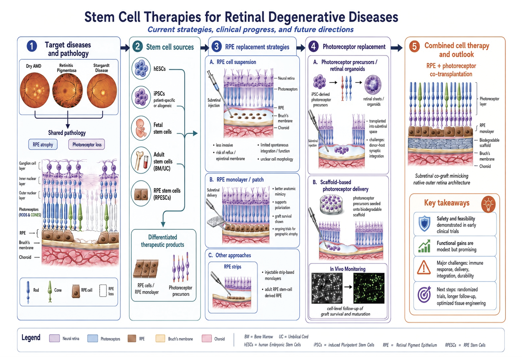 |
| Figure 1. Summary of common sources, products and delivery methods used for stem cell therapies. Cell products include suspensions of specific expanded cell populations, discrete monolayers of retinal tissue (RPE and/or PRs) to mimic native anatomy and whole slabs of retinal organoid consisting of PRs with connected interneurons. (Created with assistance from ChatGPT-5.5.) Click image to enlarge. |
RPE transplantation
By way of a quick review, the RPE serves to maintain the blood-retinal barrier and support photoreceptor function by phagocytosing used outer segments and recycling photopigment.15 The RPE has been a primary focus of cell replacement strategies due to its laminar structure and importance in defining the retinal architecture and metabolism.6 There are two main methods by which therapeutic RPE cells may be introduced to the subretinal space: injection of a cell suspension or transplantation of a cellular monolayer (with or without an artificial scaffold). The former approach is less invasive and requires a smaller retinotomy, but several clinical trials have shown that RPE cell suspensions don’t spontaneously form a normal monolayer in the subretinal space, and there’s a greater risk of reflux into the vitreous cavity. In addition, RPE cells require adhesion to a substrate to fully polarize and differentiate. Therefore, the functional status of RPE cells in suspension is unclear. Implantation of an RPE monolayer requires a more involved surgical procedure but the results more closely mimic the anatomy and physiology of the tissue to be replaced.4–6 This approach has been used in several clinical trials as well. Next, we review the results of the most recent such studies focusing on studies that have been published over the past six years. We refer the reader to our previous reviews for more comprehensive discussion of earlier landmark studies.4–6
 |
• RPE cell suspensions. In 2012 a Phase I/IIa study of subretinal injection of allogeneic hESC-RPE suspensions for treatment of advanced dry AMD and Stargardts was published.16 The four-year follow-up results for 18 patients demonstrated safety and possible efficacy in half of them.17 Since that time, several studies using hESC-RPE suspensions have reported similar results.
In 2020, a paper reported one-year results of subretinal injection of HLA-matched allogeneic iPSC-RPE cell suspension without systemic immunosuppressive medications in five patients with exudative AMD.18 There were two instances of suspected mild rejection which resolved with local administration of steroids, and all five patients developed epiretinal membranes thought to be caused by the reflux of graft cells into the vitreous space. However, the RPE graft survived in all cases, demonstrating the short-term feasibility of conservative management of a potential post-engraftment immune responses. There was neither a notable increase nor decrease in vision one year after the procedure.
In 2021, a paper reported a three-year assessment of a Phase I trial involving subretinal injection of hESC-RPE cells in three patients with SMD.19 No unexpected serious adverse events were noted during this period, and there was no evidence of abnormal proliferation or immune rejection. The rates of progression in the area of decreased autofluorescence remained within expectations of the natural course of SMD, and best corrected visual acuity improved in one patient and remained stable in the other two. Similarly, researchers reported five-year results from seven patients with SMD who underwent subretinal injection of hESC-RPE cells with no unexpected local or systemic adverse reactions.20 On average, the seven treated eyes had similar visual function and retinal sensitivity compared to the fellow eyes at five years.
A 2023 report performed a 12-month follow-up of 12 patients with advanced SMD who received hESC-RPE cells in suspension via subretinal injection.21 No unexpected surgery-related or transplantation-related adverse events occurred over this period, and interestingly, all patients had better final BCVA in operated eyes as compared to baseline.
These promising results have been tempered by several observations. First, the subretinal blebs in these cases often don’t cover the area of RPE atrophy. Second, signs of RPE reconstitution have been limited to very small regions (compared to overall bleb size) where hyper-reflective bands on OCT seem to suggest engrafted RPE. Therefore, while these studies show at least some vision improvement, the mechanisms by which subretinal injection of RPE cell suspensions work aren’t clear and is unlikely to represent direct RPE-photoreceptor mediated vision restoration.
• RPE sheets. In 2017, investigators reported the first use of an autologous iPSC-RPE monolayer after surgical removal of choroidal neovascular membrane for treatment of neovascular AMD22 and subsequent four years of follow-up23 without unexpected serious adverse events. They found that the sheet had survived in place with slight expansion of the pigmented boundaries and there was no evidence of graft rejection. Further, the thickness of the photoreceptor layer and the choroidal volume were subjectively better at the transplant site compared to non-covered areas. Visual acuity remained stable at around 20/200 over the entire duration of follow-up. Since that time, several studies using monolayers of RPE in various formations have been reported.
In 2018, one of this article’s authors (AHK) and co-workers reported preliminary results on five patients from a Phase I/IIa trial of an allogeneic, HLA-mismatched hESC-RPE sheet on a synthetic scaffold with subsequent follow up on 16 patients at one and three years.24,25 None of the subjects had unexpected serious adverse events.24 At three years, implanted eyes were more likely than non-implanted eyes to gain >5 Early Treatment Diabetic Retinopathy Study letters of BCVA and were less likely than non-implanted eyes to lose >5 letters, although this signal wasn’t statistically significant. There was no significant difference in fixation ability of either the implanted or the non-implanted eyes at this time point, but one patient demonstrated improvement in retinal sensitivity within and surrounding the area of the implant. One subject in the study passed away two years after implantation from natural causes. Histopathologic analysis of the implanted eye demonstrated survival of the graft in the subretinal space, no evidence of immune response directed at the implant, and expression of rhodopsin in the area of neurosensory retina overlying the implant.26 There were no unanticipated severe implant-related adverse events throughout the study.25
In 2024, an analysis reported a five-year follow-up of scaffold-based hESC-RPE transplantation in two patients with advanced neovascular AMD and acute subretinal hemorrhage.27 Both subjects had some extension of pigmentation beyond the original graft area and no evidence of uncontrolled RPE proliferation or immune rejection. Both subjects also had initial improvements in BCVA, fixation and retinal sensitivity, but these improvements were only sustained in one subject.
These studies suggest that surgical implantation of RPE monolayers more closely mimic the natural RPE appearance in vivo. In all these studies, the pigmentation of the implanted RPE was maintained and the OCT appearance of the implanted cells was consistent with that of RPE. One limitation of these studies is that the patient population had very advanced disease that likely put a ceiling on the potential for visual improvement. Nevertheless, both these studies and those using cell suspensions suggest that RPE transplantation is safe and feasible thereby opening the path for additional studies.
There are at least two promising ongoing studies using RPE monolayer approaches. The first is a Phase I/IIa study of induced pluripotent stem cell derived RPE on a biodegradable scaffold for geographic atrophy that’s ongoing at the National Eye Institute. Another is a Phase IIb study using human embryonic stem cell derived RPE for GA that’s sponsored by Regenerative Patch Technologies.
• Other RPE replacement strategies. An alternative, intermediate approach between suspensions and sheets involves RPE cells grown in monolayers within narrow grooves. These “RPE strips” were successfully injected into the subretinal space of three patients (one with advanced dry AMD and two with RP) using a 31G cannula. A paper described safety and efficacy outcomes of this Phase I/II trial in 2025.28 No unexpected serious adverse events were noted, but a suspected immune reaction and epiretinal membrane formation were observed in one patient each. At the one-year mark, there was a reduction in the abnormal area in all three patients, with one patient also reporting significant vision-related quality of life improvement.
Another option being explored in RPE transplantation is the direct use of adult RPE stem cells isolated from post-mortem tissue and expanded in culture. A 2025 paper reported interim results from the first-in-human study of RPESC-RPE cells delivered as a suspension via subretinal injection in six patients with dry AMD.29 The treatment was well-tolerated with no cell-product-related severe adverse events. Treated eyes had improvements in BCVA, whereas untreated eyes had reduced visual acuity compared to baseline consistent with the natural history of GA. These results suggest that there may be advantages to using this cell type although this method would have all the limitations of cell suspension methods listed above.
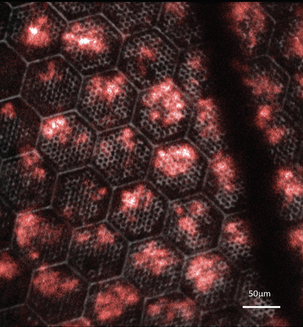 |
| Figure 2. Adaptive optics scanning light ophthalmoscope image of a honeycomb scaffold seeded with photoreceptor precursors (red) in the subretinal space of a non-human primate model in vivo. Infrared reflectance image of the scaffold microstructure is overlaid with the false-colored fluorescence image of photoreceptor precursors expressing tdTomato. |
Photoreceptor transplantation
The results of RPE transplantation studies described above raise the obvious question of whether a similar approach is viable for photoreceptors as well. Photoreceptor transplantation faces many of the same challenges as RPE transplantation, such as stringent organization and polarity demands. Photoreceptors come in varied types, and they must also functionally integrate with host tissue for proper transduction of visual signals.30 Photoreceptor replacement is still being optimized at the pre-clinical stage. One line of investigation in photoreceptor transplantation is based on human pluripotent stem cell-derived photoreceptor precursors (hPSC-PRPs). These cells are produced within three-dimensional retinal organoids that recapitulate human retinogenesis. Within this category, two main products have shown promise: iPSC-derived retinal sheets31,32 and suspensions of photoreceptor precursors.33,34
In similar fashion to RPE suspensions, direct subretinal injections of purified photoreceptor precursor cells have been associated with inconsistent delivery and reflux into the vitreous cavity, but without the prevalence of epiretinal membrane formation. Better anatomic success has been seen with the use of structured retinal sheets, but synaptic connection has remained limited due to the presence of interneurons and the formation of rosettes. Efforts are underway to use genome editing to optimize donor-host connectivity by depleting the interneurons.35
Another method being explored by one of this article’s authors (JEM) is the use of synthetic scaffolds seeded with isolated PRPs.36 To track the survival, distribution, and morphology of transplanted photoreceptors in animal models in vivo, hPSC-PRPs can be genetically engineered to express fluorescent reporter proteins. Pairing this approach with adaptive optics scanning laser ophthalmoscopy, it’s possible to assess both the microstructure of the scaffold and the survival and maturation of the photoreceptor transplant at a cellular level in the living eye (Figure 2).
Photoreceptor cell replacement therapies are now being evaluated in a small number of clinical trials. In 2023, two-year results from the first in-human study of retinal organoid transplantation were published.37 Two subjects with RP were implanted with allogeneic iPSC-derived retinal organoids, which survived with neither immune rejection nor unexpected overgrowth. Visual acuity and visual field both remained stable, but retinal sensitivity decreased across the entire measurement area, including at the points overlying the local transplant site.
Adaptive optics optical computed tomography was performed on one patient who received retinal organoid transplantation.38 It was observed that the implanted region had outer plexiform layer-like lines and reflective particles consistent in appearance with outer segment discs, suggesting that the graft may have developed functional morphology. Another clinical trial which is now underway is the Phase I/IIa CLARICO trial for OpCT-001, the first iPSC-derived cell therapy for primary photoreceptor diseases.
Because of the inherent interdependence of RPE and photoreceptor cells and their concomitant loss in advanced disease, researchers are also investigating co-transplantation of both cell types.39 In a proof-of-concept study involving an immunodeficient laser induced rat model of advanced retinal degeneration researchers demonstrated safe transplantation of a co-graft consisting of a retinal organoid plus a polarized RPE monolayer on a parylene scaffold.40 Immunohistochemistry revealed that these co-grafts generated photoreceptors and developed neuronal processes which migrated into the host retina. Visual function assessments using optokinetic nystagmus showed improvement in the implanted animals compared to controls. Further optimization of the co-grafting process is still required, but these results support the translational potential of this technique.
Conclusions and future directions
These recent clinical trials contribute to a growing body of evidence pointing to the safety and feasibility of stem cell therapies when performed with appropriate oversight, expertise and expectation. Overall, well-performed cell-based interventions have been associated with reasonable serious adverse event rates for clinical implementation. Continued research into alternative immunosuppression regimens and modes of delivery may further help reduce risks. Functional improvement has been difficult to demonstrate, partially owing to an inherent limitation of early phase studies in a relatively small sample size and often advanced disease. Efficacy analysis is further complicated by significant heterogeneity in control groups and outcome measures.
However, improvements in visual acuity, visual field and retinal sensitivity have been noted in preliminary studies, especially those implementing careful patient selection. Further, a recent meta-analysis found that, across a total of 18 studies describing quantitative data from 224 eyes, cell therapy was associated with significantly improved BCVA in AMD, RP and SMD.41
Ultimately, randomized controlled trials with longer follow-up will be critical to establish definitive therapeutic benefit, assess treatment durability and monitor for late-onset or rare adverse effects. It’s clear that, with continued optimization of tissue engineering and graft delivery, stem cells hold tremendous therapeutic potential in retinal degenerative diseases. It’s important to note that poorly done studies (often prematurely claiming effectiveness) have had catastrophic results and we encourage readers to review an NEJM article summarizing these events.42 Physicians and patients should exercise caution when considering enrolling in clinical trials that seem to promise or exaggerate positive results, especially if payment for participation is required.42 RS
REFERENCES
1. Wert KJ, Lin JH, Tsang SH. General pathophysiology in retinal degeneration. Dev Ophthalmol 2014;53:33.
2. Liu W, Liu S, Li P, Yao K. Retinitis pigmentosa: Progress in molecular pathology and biotherapeutical strategies. Int J Mol Sci 2022;23:9:4883.
3. Girgis S, Lee LR. Treatment of dry age-related macular degeneration: A review. Clin Exp Ophthalmol 2023;51:8:835-852.
4. Kashani AH. Stem cell therapy in nonneovascular age-related macular degeneration. Invest Ophthalmol Vis Sci 2016;57:5:ORSFm1-ORSFm9.
5. Nazari H, Zhang L, Zhu D, et al. Stem cell based therapies for age-related macular degeneration: The promises and the challenges. Prog Retin Eye Res 2015;48:1-39.
6. Kashani AH. Stem cell-derived retinal pigment epithelium transplantation in age-related macular degeneration: Recent advances and challenges. Curr Opin Ophthalmol 2022;33:3:211-218.
7. Long J, Xu Z, Hu P, Ye Y, Long D. Stem cell therapy for inherited retinal diseases: Trends and insights from 2000 to 2024. Cell Transplant 2025;34.
8. Nair DSR, Thomas BB. Stem cell-based treatment strategies for degenerative diseases of the retina. Curr Stem Cell Res Ther 2022;17:3:214-225.
9. Assen LS, Jongsma KR, Isasi R, Tryfonidou MA, Bredenoord AL. Recognizing the ethical implications of stem cell research: A call for broadening the scope. Stem Cell Reports 2021;16:7:1656.
10. Singh MS, Park SS, Albini TA, et al. Retinal stem cell transplantation: Balancing safety and potential. Prog Retin Eye Res. Mar 2020;75.
11. Hoch AI, Leach JK. Concise review: Optimizing expansion of bone marrow mesenchymal stem/stromal cells for clinical applications. Stem Cells Transl Med 2014;3:5:643-652.
12. Cerneckis J, Cai H, Shi Y. Induced pluripotent stem cells (iPSCs): Molecular mechanisms of induction and applications. Signal Transduction and Targeted Therapy 2024;9:1:112.
13. Salero E, Blenkinsop TA, Corneo B, et al. Adult human RPE can be activated into a multipotent stem cell that produces mesenchymal derivatives. Cell Stem Cell 2012;10:1:88-95.
14. Stern JH, Saini JS, Temple S, et al. Human retinal pigment epithelium stem cell (RPESC). Adv Exp Med Biol 2015;854:557-562.
15. George SM, Lu F, Rao M, Leach LL, Gross JM. The retinal pigment epithelium: Development, injury responses, and regenerative potential in mammalian and non-mammalian systems. Prog Retin Eye Res 2021;85.
16. Schwartz SD, Hubschman JP, Heilwell G, et al. Embryonic stem cell trials for macular degeneration: A preliminary report. Lancet 2012;379:9817:713-720.
17. Schwartz SD, Tan G, Hosseini H, Nagiel A. Subretinal transplantation of embryonic stem cell-derived retinal pigment epithelium for the treatment of macular degeneration: An assessment at 4 years. Invest Ophthalmol Vis Sci 2016;57:5:ORSFc1-ORSFc9.
18. Sugita S, Mandai M, Hirami Y, et al. HLA-matched allogeneic iPS cells-derived RPE transplantation for macular degeneration. J Clin Med 2020;9:7:2217.
19. Sung Y, Lee MJ, Choi J, et al. Long-term safety and tolerability of subretinal transplantation of embryonic stem cell-derived retinal pigment epithelium in Asian Stargardt disease patients. Br J Ophthalmol 2021;105:6:829-837.
20. Li SY, Liu Y, Wang L, et al. A phase I clinical trial of human embryonic stem cell-derived retinal pigment epithelial cells for early-stage Stargardt macular degeneration: 5-years’ follow-up. Cell Prolif 2021;54:9.
21. Fernandes RAB, Lojudice FH, Zago Ribeiro L, et al. Transplantation of subretinal stem cell-derived retinal pigment epithelium for Stargardt disease: A phase I clinical trial. Retina 2023;43:2:263-274.
22. Mandai M, Watanabe A, Kurimoto Y, et al. Autologous induced stem-cell-derived retinal cells for macular degeneration. N Engl J Med 2017;376:11:1038-1046.
23. Takagi S, Mandai M, Gocho K, et al. Evaluation of transplanted autologous induced pluripotent stem cell-derived retinal pigment epithelium in exudative age-related macular degeneration. Ophthalmol Retina 2019;3:10:850-859.
24. Kashani AH, Lebkowski JS, Rahhal FM, et al. One-year follow-up in a phase 1/2a clinical trial of an allogeneic RPE cell bioengineered implant for advanced dry age-related macular degeneration. Transl Vis Sci Technol 2021;10:10.
25. Humayun MS, Clegg DO, Dayan MS, et al. Long-term follow-up of a phase 1/2a clinical trial of a stem cell-derived bioengineered retinal pigment epithelium implant for geographic atrophy. Ophthalmology 2024;131:6:682-691.
26. Kashani AH, Lebkowski JS, Hinton DR, et al. Survival of an HLA-mismatched, bioengineered RPE implant in dry age-related macular degeneration. Stem Cell Reports 2021;17:3:448-458.
27. da Cruz L, Soomro T, Georgiadis O, Nommiste B, Sagoo MS, Coffey P. The fate of RPE cells following hESC-RPE patch transplantation in haemorrhagic wet AMD: Pigmentation, extension of pigmentation, thickness of transplant, assessment for proliferation and visual function-A 5 year-follow up. Diagnostics (Basel) 2024;14:10.
28. Sakai D, Mandai M, Hirami Y, et al. Transplant of induced pluripotent stem cell-derived retinal pigment epithelium strips for macular degeneration and retinitis pigmentosa. Ophthalmology Science 2025;5:4.
29. Rao RC, Arduini BL, Borden S, et al. Safety and tolerability of RPESC-RPE transplantation in patients with dry age-related macular degeneration: Low-dose clinical outcomes. Cell Stem Cell 2025;32:11:1659-1670.e4.
30. Monville C, Goureau O, M’Barek K Ben. Photoreceptor cell replacement using pluripotent stem cells: Current knowledge and remaining questions. Cold Spring Harb Perspect Med 2023;13:2:a041309.
31. Shirai H, Mandai M, Matsushita K, et al. Transplantation of human embryonic stem cell-derived retinal tissue in two primate models of retinal degeneration. Proc Natl Acad Sci U S A 2016;113:1:E81-E90.
32. Seiler MJ, Aramant RB. Cell replacement and visual restoration by retinal sheet transplants. Prog Retin Eye Res 2012;31:6:661-687.
33. Aboualizadeh E, Phillips MJ, McGregor JE, et al. Imaging transplanted photoreceptors in living nonhuman primates with single-cell resolution. Stem Cell Reports 2020;15:2:482-497.
34. Gasparini SJ, Tessmer K, Reh M, et al. Transplanted human cones incorporate into the retina and function in a murine cone degeneration model. J Clin Invest 2022;132:12.
35. Watanabe M, Yamada T, Koike C, Takahashi M, Tachibana M, Mandai M. Transplantation of genome-edited retinal organoids restores some fundamental physiological functions coordinated with severely degenerated host retinas. Stem Cell Reports 2025;20:2:102393.
36. Lee IK, Xie R, Luz-Madrigal A, et al. Micromolded honeycomb scaffold design to support the generation of a bilayered RPE and photoreceptor cell construct. Bioact Mater 2024;30:142.
37. Hirami Y, Mandai M, Sugita S, et al. Safety and stable survival of stem-cell-derived retinal organoid for 2 years in patients with retinitis pigmentosa. Cell Stem Cell 2023;30:12:1585-1596.e6.
38. Ishikura M, Muraoka Y, Hirami Y, Tu HY, Mandai M. Adaptive optics optical coherence tomography analysis of induced pluripotent stem cell-derived retinal organoid transplantation in retinitis pigmentosa. Cureus 2024;16:7.
39. Rajendran Nair DS, Seiler MJ, Martinez-Camarillo JC, et al. Stem cell-derived co-grafts contribute to retinal reconstruction and visual functional improvement in a laser damaged rat model. Mil Med Res 2025;12:1:23.
40. Thomas BB, Lin B, Martinez-Camarillo JC, et al. Co-grafts of human embryonic stem cell derived retina organoids and retinal pigment epithelium for retinal reconstruction in immunodeficient retinal degenerate Royal College of Surgeons rats. Front Neurosci 2021;15.
41. Khaboushan AS, Ebadpour N, Moghadam MMJ, Rezaee Z, Kajbafzadeh AM, Zolbin MM. Cell therapy for retinal degenerative disorders: A systematic review and three-level meta-analysis. J Transl Med 2024;22:1.
42. Kuriyan AE, Albini TA, Townsend JH, et al. Vision loss after intravitreal injection of autologous “stem cells” for AMD. N Engl J Med 2017;376:11:1047-1053.