Going from bench to bedside is an incredibly long process with many regulatory hurdles, all intended to ensure safety and efficacy of new therapeutics before they are widely adopted in medicine. In the retina space, it probably wasn’t until brolucizumab that many of us realized clinical trial safety data doesn’t always translate into broader patient safety once you go from hundreds of patients being treated in trials to many thousands in the real world. We’ve seen this repeated to some extent with the complement inhibitors and their (fortunately rarer) occurrence of occlusive retinal vasculitis compared to brolucizumab.
At the recent Retina Society meeting in Los Angeles, our colleague Dr. David Brown gave a striking presentation on the association between anti-complement therapy and thickening of the peripapillary retinal nerve fiber layer.
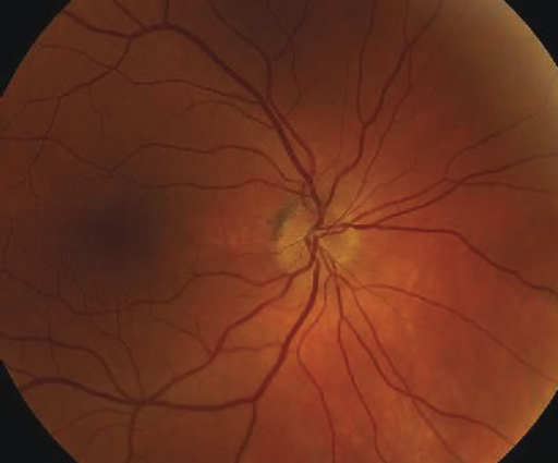 |
| Image courtesy of Flanary WE, et al./eyerounds.org2 |
First off, I have to give him kudos for actually getting peripapillary RNFL scans on so many patients for so long, as it’s something that I rarely order. During his presentation, he reported performing serial RNFL scans on treatment naïve eyes, anti-VEGF injected eyes, and anti-complement injected eyes (both pegcetacoplan and avacincaptad pegol).
The treatment naïve and anti-VEGF treated eyes had a slight annual decrease in thickening as would be expected for aging, while the anti-complement treated eyes had an annual 4 to 5 µm increase. He hypothesized that this isn’t a neuroprotective reversal of RNFL loss but rather a thickening of the existing RNFL.
So what’s the big deal? There appears to be a <1-percent risk of acute ischemic optic neuropathy in patients receiving anti-complement inhibitors based on data from the pivotal trials. This progressive RNFL thickening may be a mechanism similar to how a crowded optic disc with a small cup is considered a “disc at risk” for non-arteritic ischemic optic neuropathies. Dave showed a striking example where you could see progressive, dramatic shrinking of the cup on serial imaging.
I walked away with a couple of take-home messages: 1) In patients receiving anti-complement injections, it may be worthwhile to obtain peripapillary RNFL thickness measurements, with a plan to hold injections if there is a noticeable increase above the normative threshold. 2) Use caution in treating patients who already have a small cup-to-disc ratio <0.2 as RNFL thickening in these eyes may be more likely to choke the disc. RS
|
1. Brown, DM (2026, September 25). Anti-Complement Therapy Induced Thickening of the Retinal Nerve Fiber Layer: A Longitudinal Analysis [Paper presentation]. 59th Annual Meeting of The Retina Society, Los Angeles, CA.
2. Image: Flanary WE, Phillips D, Kardon RH. Posterior ischemic optic neuropathy. April 3, 2015; Available from: https://eyerounds.org/cases/212-PION.htm. Used under Creative Commons License: https://creativecommons.org/licenses/by-nc-nd/3.0/.