Take-home points
|
 |
Bios Dr. Kuriyan is on the Retina Service at Wills Eye Hospital (and also practices at Mid Atlantic Retina) and is an associate professor of ophthalmology at Sidney Kimmel Medical College of Thomas Jefferson University. |
Proliferative vitreoretinopathy remains the most common and formidable cause of surgical failure in the treatment of rhegmatogenous retinal detachment.1,2 Characterized by the growth and contraction of fibrocellular membranes on both the retinal surfaces and within the vitreous cavity, PVR represents an exaggerated, protracted wound-healing response. Retinal pigment epithelial cells, glial cells, and inflammatory macrophages undergo epithelial-mesenchymal transition mediated by various cytokines and growth factors.3,4 This multifactorial fibroproliferative process mechanically distorts the retina, often inducing new retinal breaks or reopening existing ones.5 Despite major advances in vitreoretinal surgical techniques and instrumentation, preventing and managing severe PVR continues to challenge retina specialists worldwide. In this article, we outline novel and emerging approaches to diagnosing and managing PVR, highlighting advancements in clinical imaging, surgical techniques, and targeted pharmacotherapy.
1) Diagnosing PVR clinical criteria and pvr scoring
The foundation of PVR management begins with accurate clinical diagnosis and standardized grading. The first widely recognized classification system was published by The Retina Society Terminology Committee in 1983.6 This system categorized PVR based on clinical signs and geographic distribution into four progressive grades:
• Grade A: Vitreous haze and vitreous pigment clumps. This is the earliest clinical manifestation of the disease process.
• Grade B: Surface retinal wrinkling, rolled edges of retinal breaks, retinal stiffness and vessel tortuosity.
• Grade C: Full-thickness, fixed retinal folds. This was historically subdivided by the number of quadrants involved (C-1 to C-3).
• Grade D: Fixed retinal folds in all four quadrants forming a funnel detachment (D-1 wide, D-2 narrow, D-3 closed).
While the 1983 Retina Society classification laid the necessary groundwork, it was later updated to better account for anterior PVR and the exact types of contraction.7 Using a standard PVR scoring system is helpful in practice. Not only does it dictate the surgical approach, such as anticipating the need for an encircling scleral buckle, extensive membrane peeling or retinectomy, but it also standardizes patient cohorts for clinical trials and retrospective studies.
Imaging the PVR interface
While PVR has historically been a clinical diagnosis, advanced optical coherence tomography imaging has revolutionized our ability to identify its earliest microstructural precursors. The onset of PVR is marked by the intraretinal migration of macrophages, glial cells, and transdifferentiated RPE cells, which disrupt the retinal microarchitecture, and can appear as outer retinal hyperreflective dots on OCT before overt clinical signs appear.8
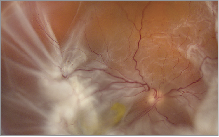 |
| The contractile membranes of PVR result in generalized retinal shrinkage, fixed retinal folds, and characteristic "star folds," which are most evident from 4:00 to 11:00 in this photograph of a retinal detachment involving the macula. Photo: Jesse Vislisel, MD/Brice Critser, CRA/eyerounds.org |
OCT findings and retinal reattachment physiology
Recent investigations by Muni and colleagues have utilized high-resolution OCT to characterize the in vivo physiology of retinal detachment and reattachment. Muni et al. developed a morphologic grading system that classifies RRD based on progressive OCT imaging features, ranging from neurosensory separation to the complete loss of photoreceptor inner and outer segments.9
Following procedures such as pneumatic retinopexy, normal retinal reattachment occurs in microstructural stages. Delayed progression through these reattachment stages is associated with postoperative anatomic abnormalities, including the formation of outer retinal folds, persistent subfoveal fluid blebs, and early epiretinal membrane formation.10 Identifying these early structural anomalies provides a window into the retina’s mechanical stiffness and early cellular proliferation, alerting the surgeon to an increased risk of clinically significant PVR before macroscopic fixed folds appear.
Reflectivity in PVR
The optical properties of the detached retina and overlying membranes serve as profound biomarkers for PVR. Histopathological changes, such as the intraretinal migration of RPE cells and disorganized retinal layers, increase the backscatter of light on OCT.11
In a recent study by our group, adjusted retinal pixel intensity (ARPI) was evaluated to quantify this hyperreflectivity and correlate it with the presence of PVR and postoperative outcomes.12 ARPI calculates the difference between the mean pixel intensity of the detached neurosensory retina and the region of the vitreous directly overlying it. It was found that an increased ARPI was independently associated with the presence of Grade C PVR and lower rates of single-surgery anatomic success (SSAS). Notably, this trend was observed even in patients who didn't have clinically apparent PVR at the time of initial presentation, suggesting that quantifying retinal hyperintensity may serve as a valuable, non-invasive imaging biomarker to aid in preoperative risk stratification.13 By identifying high-risk eyes prior to surgery, retina specialists can potentially proactively employ surgical techniques such as internal limiting membrane peeling, or prophylactic pharmacotherapy, to optimize outcomes and prevent surgical failure.
2) Managing PVR surgical techniques
The surgical management of PVR requires meticulous relief of all mechanical traction. Once core and peripheral vitrectomies are complete, surgeons employ advanced techniques to ensure long-term anatomic success.
• Internal limiting membrane peeling. The ILM acts as a primary scaffold for cellular proliferation in PVR. A robust body of literature supports the utility of ILM peeling in both primary high-risk RRDs and complex PVR cases.14 Peeling the ILM effectively removes the substrate upon which myofibroblasts and RPE cells migrate and contract.
In cases of uncomplicated retinal detachments, Shah and colleagues reported ILM peeling decreases rates of postoperative ERM formation.15 A small retrospective study of patients with Group B PVR found a higher single surgery success rate and less ERM formation in patients who underwent ILM peeling compared to patients who did not.16 Furthermore, a study by Wakabayashi and colleagues also demonstrates that primary ILM peeling during RRD with Grade C PVR repair reduces the incidence of retinal re-detachment and postoperative PVR.17 These studies support that ILM peeling in RD surgery with or without PVR may improve anatomical outcomes.
• PFO-guided retinectomy. In cases of advanced, severe Grade C PVR with intrinsic retinal shortening, retinectomy is often necessary.18 Historically, patients requiring retinectomy for Grade C PVR have faced guarded outcomes, with SSAS rates hovering between 61 and 67 percent at six months.19 Surgical failure in these complex cases is frequently caused by new fibrosis, missed fibrosis during the initial operation or intrinsic fibrosis and immature pre-retinal membranes that are difficult to visualize.
To combat these challenges, our team recently evaluated outcomes utilizing a novel Perfluoro-n-Octane (PFO)-guided retinectomy technique.20 This technique leverages the identification of a deformation of the PFO bubble to visually identify the ideal posterior edge of the retinectomy, ensuring that all immature membranes and intrinsic fibrosis are adequately removed. Our data demonstrated that patients undergoing PFO-guided retinectomy achieved nearly an 89 percent SSAS rate at six months, representing a significant improvement over institutional historical controls and the pooled historical controls used in recent major trials. This technique offers a potential effective strategy to improve surgical outcomes in eyes with Grade C PVR.
 |
Medications: From past failures to promising horizons
While surgery expertly addresses the mechanical forces of PVR, resolving the underlying cellular proliferation requires targeted pharmacotherapy.
• Past therapeutics. Historically, numerous antimetabolites and anti-inflammatory agents have been investigated with underwhelming results. 5-fluorouracil (5-FU) and low-molecular-weight heparin (LMWH) were in the landmark PRIVENT trial.21 However, the findings were unable to be replicated in a subsequent study.22 Similarly, while intraocular corticosteroids effectively reduce postoperative inflammation, large-scale randomized trials have consistently failed to show a definitive reduction in overall PVR rates.23
• Tumor necrosis factor inhibitors. Recent translational research has focused on the precise signaling cascades driving EMT in PVR. Cigarette smoke is an established, modifiable risk factor for PVR.24 Laboratory research has elucidated that cigarette smoke extract significantly upregulates pro-inflammatory cytokines, specifically tumor necrosis factor-alpha (TNF-α), driving the EMT of RPE cells via the NF-κB/Snail signaling axis. In murine models of PVR, inhibiting TNF-α or the downstream NF-κB pathway blocks this transition and significantly reduces PVR severity.25 These findings position TNF inhibitors as highly promising, targeted prophylactic agents for patients at high risk of PVR, particularly those with a history of smoking or chronic inflammation.
A small study of 60 patients examined the use of an intravitreal anti-TNF-α agent, infliximab, to inhibit PVR formation in patients with RRDs with Grade C PVR. There was a non-significant improved single surgery success rate in the infliximab group (26/30) compared to the control group (23/30). The treatment group had slightly better visual outcomes (Snellen equivalent ≈ 20/180) compared to the control group (Snellen equivalent ≈ 20/280; p=0.044).26
• Methotrexate. While targeting specific signaling pathways like TNF offers a promising prophylactic strategy, combating the broader fibrocellular proliferation characteristic of established EMT requires potent antimetabolite therapy. In this regard, perhaps the most exciting development in the medical management of PVR is the use of intravitreal methotrexate. As an antimetabolite, methotrexate effectively inhibits the rapid proliferation of RPE and glial cells.
The Phase III GUARD trial provided encouraging evidence for adjunctive intravitreal methotrexate using ADX-2191. While eyes receiving ADX-2191 had a 24-percent rate of recurrent RD requiring reoperation compared to 39 percent in historical controls, the randomized comparison with routine surgical care didn’t demonstrate a statistically significant difference (18.8 vs. 20.6 percent).27 Therefore, these findings should be interpreted as promising rather than definitive, especially given the trial’s substantial treatment burden of 13 injections over 16 weeks.
Corroborating this, early data from the FIXER trial, presented at ASRS, also support investigation of intraoperative methotrexate infusion followed by postoperative injections for PVR prevention.28 Together, these data suggest that methotrexate may play an important adjunctive role for complex RDs and RDs with PVR.
Conclusion
The emerging approach to PVR is shifting from treating established mechanical contraction toward earlier identification of biologic risk and more targeted intervention. Quantitative imaging, refined surgical techniques, and adjunctive pharmacotherapy may ultimately allow treatments that improve visual and surgical outcomes in patients with PVR. Continued collaboration and rigorous clinical trials will be vital as we refine these techniques and integrate them into standard clinical practice to ultimately improve patient outcomes. RS
REFERENCES
1. Kwon OW, Song JH, Roh MI. Retinal detachment and proliferative vitreoretinopathy. Dev Ophthalmol. 2016;55:154-162.
2. Idrees S, Sridhar J, Kuriyan AE. Proliferative vitreoretinopathy: a review. Int Ophthalmol Clin. 2019;59(1):221-240.
3. Hiscott PS, Grierson I, McLeod D. Retinal pigment epithelial cells in epiretinal membranes: an immunohistochemical study. Br J Ophthalmol. 1984;68(10):708-715.
4. Shahlaee A, Woeller CF, Philp NJ, et al. Translational and clinical advancements in management of proliferative vitreoretinopathy. Curr Opin Ophthalmol. 2022;33(3):219-227.
5. Kiss CG, Richter-Müksch S, Sacu S, et al. Anatomy and function of the macula after surgery for retinal detachment complicated by proliferative vitreoretinopathy. Am J Ophthalmol. 2007;144(6):872-877.
6. The Retina Society Terminology Committee. The classification of retinal detachment with proliferative vitreoretinopathy. Ophthalmology. 1983;90(2):121-125.
7. Machemer R, Aaberg TM, Freeman HM, et al. An updated classification of retinal detachment with proliferative vitreoretinopathy. Am J Ophthalmol. 1991;112(2):159-165.
8. Jhaveri A, Martins Melo I, Pecaku A, et al. Outer retinal hyperreflective dots: a potential imaging biomarker in rhegmatogenous retinal detachment. Ophthalmol Retina. 2023;7(12):1087-1096.
9. Martins Melo I, Bansal A, Naidu S, et al. Morphologic stages of rhegmatogenous retinal detachment assessed using swept-source OCT. Ophthalmol Retina. 2023;7(5):398-405.
10. Muni RH, Darabad MN, Oquendo PL, et al. Outer retinal corrugations in rhegmatogenous retinal detachment: the retinal pigment epithelium–photoreceptor dysregulation theory. Am J Ophthalmol. 2023;245:14-24.
11. Pastor JC, Méndez MC, de la Fuente MA, et al. Intraretinal immunohistochemistry findings in proliferative vitreoretinopathy with retinal shortening. Ophthalmic Res. 2006;38(4):193-200.
12. Cheng BT, Yu MD, Kim ED, Yonekawa Y, Hsu J, Kuriyan AE. Retinal pixel intensity on OCT correlates with proliferative vitreoretinopathy in patients with macula-off retinal detachment. In Press.
13. Ferro Desideri L, Danilovska T, Bernardi E, et al. Artificial intelligence-enhanced OCT biomarkers analysis in macula-off rhegmatogenous retinal detachment patients. Transl Vis Sci Technol. 2024;13(10):21.
14. Fallico M, Russo A, Longo A, et al. Internal limiting membrane peeling versus no peeling during primary vitrectomy for rhegmatogenous retinal detachment: a systematic review and meta-analysis. PLoS One. 2018;13(7):e0201010.
15. Rao RC, Blinder KJ, Smith BT, et al. Internal limiting membrane peeling for primary rhegmatogenous retinal detachment repair. Ophthalmology. 2013;120(5):1102-1103.
16. Rao RC, Shah GK. Internal limiting membrane peeling in macula-off retinal detachment complicated by grade B proliferative vitreoretinopathy. Am J Ophthalmol. 2018;196:212-213.
17. Wakabayashi T, Samuelson AG, Oshima Y, et al. Internal limiting membrane peeling for grade C proliferative vitreoretinopathy: an international multicenter study. Ophthalmol Retina. 2025.
18. Mahmoudzadeh R, et al. Outcomes of retinectomy for proliferative vitreoretinopathy. Retina. 2023.
19. Banerjee PJ, Xing W, Bunce C, et al. Surgical outcomes for rhegmatogenous retinal detachment complicated by proliferative vitreoretinopathy. Am J Ophthalmol. 2017.
20. Kuriyan AE, Zhang J, Yonekawa Y, et al. Novel technique of perfluoro-n-octane (PFO)-guided retinectomy improves single-surgery anatomic success (SSAS) in eyes with proliferative vitreoretinopathy (PVR). Presented at: American Society of Retina Specialists (ASRS) Annual Meeting; August 2025.
21. Asaria RH, Kon CH, Bunce C, et al. Adjuvant 5-fluorouracil and heparin prevents proliferative vitreoretinopathy: results from a randomized, double-blind, controlled clinical trial. Ophthalmology. 2001;108(7):1179-1183.
22. Charteris DG, Aylward GW, Wong D, et al. A randomized controlled trial of combined 5-fluorouracil and low-molecular-weight heparin in management of established proliferative vitreoretinopathy. Ophthalmology. 2004;111(12):2240-2245.
23. Schaub F, Adler W, Enders P, et al. Intravitreal dexamethasone implant for the prevention of proliferative vitreoretinopathy: The VIPER Study. Am J Ophthalmol. 2022;239:150-161.
24. Eliott D, Stryjewski TP, Andreoli MT, et al. Smoking is a risk factor for proliferative vitreoretinopathy after traumatic retinal detachment. Retina. 2017;37(7):1229-1235.
25. Wang V, Heffer A, Roztocil E, et al. TNF-α and NF-κB signaling play a critical role in cigarette smoke-induced epithelial-mesenchymal transition of retinal pigment epithelial cells in proliferative vitreoretinopathy. PLoS One. 2022;17(9):e0271950.
26. Younes A, Hamza H, Omar H, et al. Intravitreal infliximab for the treatment of proliferative vitreoretinopathy (FIXER). Ophthalmol Retina. 2024;8:1044-1051.
27. Aldeyra Therapeutics. ADX-2191 Phase 3 GUARD Trial Results. Presented at: American Academy of Ophthalmology (AAO) Annual Meeting; 2023.
28. Riemann CD. FIXER Trial: Intraoperative and postoperative methotrexate for PVR prevention. Presented at: American Society of Retina Specialists (ASRS) Annual Meeting; 2024.