Take-home points
|
 |
Bios Dr. Acabá-Berrocal practices in the Department of Ophthalmology, Wills Eye Hospital, Thomas Jefferson University, in Philadelphia. |
Surgical management of complications related to diabetic retinopathy remains challenging due to the heterogeneity of the disease. No two cases present in exactly the same way. The most common pathologies requiring surgical intervention include vitreous hemorrhage, tractional retinal detachment and combined tractional–rhegmatogenous retinal detachment.
In many diabetic eyes, the posterior hyaloid is partially attached and thickened, creating a scaffold for neovascularization. These neovascular complexes often form strong adhesions to the retina, making their removal technically demanding.
Complications during diabetic vitreoretinal surgery can be broadly classified into intraoperative and postoperative complications.
Intraoperative complications occur in approximately 13 to 30 percent of cases and include:
• bleeding during fibrovascular tissue dissection;
• iatrogenic retinal breaks caused by strong fibrovascular adhesions to a thinned retina;
• poor intraoperative visualization due to prolonged surgery and elevated intraocular pressure in phakic eyes; and
• lens trauma and cataract formation.1-5
Postoperative complications include:
• early or late postoperative vitreous hemorrhage (20 to 32 percent), the most common complication;
• cataract progression (18 to 21 percent) within one year;
• elevated intraocular pressure (~10 percent);
• neovascular glaucoma (5 to 9 percent); and
• rhegmatogenous retinal detachment.
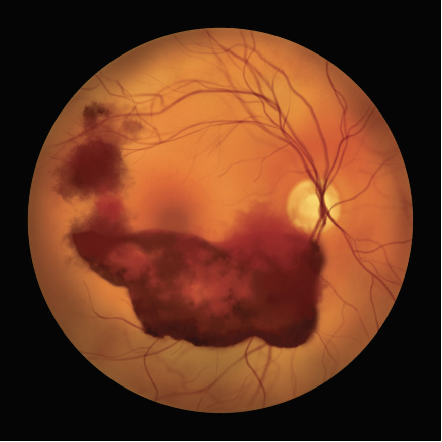 |
| Vitreous hemorrhage is one of the most common pathologies requiring surgical intervention in DR patients. Photo: Getty. |
Intraoperative pitfalls and their prevention
There are several issues to watch out for during surgery:
• Poor intraoperative visualization. Poor visualization is common in diabetic eyes and may result from:
• inadequate pupillary dilation;
• corneal epithelial instability;
• intraoperative bleeding;
• posterior synechiae; and
• lens opacities.
Maintaining adequate dilation throughout surgery is essential. Intracameral epinephrine and glucose-containing irrigation solutions may assist in maintaining dilation in phakic eyes.
Corneal clarity can be improved by topical glycerine, which helps reduce corneal edema. Mechanical corneal scraping should be avoided when possible, as diabetics are prone to persistent epithelial defects.
Iris hooks may be useful when dilation is inadequate. However, controlling intraoperative bleeding remains the most critical factor for maintaining good visibility.
Clear visualization of the peripheral retina during membrane dissection is essential because inadvertent traction in the periphery may occur during manipulation. Digital 3D visualization systems can improve magnification while maintaining a wide field of view, unlike traditional macular lenses used with standard microscope viewing.
• Lens trauma in phakic eyes. In phakic patients, inadvertent trauma to the lens can significantly impair visualization and may lead to rapid progression to a white cataract in the early postoperative period.
This complication can be minimized by avoiding crossing instruments across the visual axis toward the contralateral periphery and maintaining awareness of the posterior capsule location throughout surgery.
• Intraoperative bleeding. Intraoperative bleeding is the most frequent complication during diabetic vitrectomy. It typically occurs during segmentation or peeling of fibrovascular membranes and detachment of the posterior hyaloid. Bleeding significantly compromises visualization and must be controlled promptly.
Key strategies to minimize bleeding include:
• ensuring adequate systemic blood pressure control during surgery;
• temporarily raising intraocular pressure during vessel segmentation;
• cauterizing vessels using endodiathermy or laser when necessary; and
• avoiding sudden IOP fluctuations, particularly hypotony.
Large blood clots forming over detached retina are difficult to remove and may lead to iatrogenic retinal breaks or hinder further membrane dissection.
• Iatrogenic retinal breaks. Avoiding iatrogenic retinal breaks during hyaloid removal and fibrovascular membrane dissection is critical, as such breaks are associated with a 2.5 to 3.9-fold increased risk of poor visual outcomes.8
If a break occurs, the surgeon must relieve surrounding traction, apply adequate laser treatment and consider long-term tamponade agents.
Although not always preventable, several strategies can reduce the risk:
• Maintain optimal visualization at all times. Tissue removal should never be attempted when visualization is poor.
• Preoperative panretinal photocoagulation. Peripheral PRP strengthens retinal adhesions and reduces traction-related breaks during surgery.
• Early removal of peripheral vitreous. Clearing vitreous adhesions at the beginning of the case reduces traction generated during intraocular maneuvers.
• Identify a safe dissection plane. Whenever possible, locate the plane between retina and fibrovascular tissue before initiating dissection.
• Initiate dissection near the optic nerve. If a plane isn’t easily identifiable, detaching the hyaloid around the optic disc and progressing toward the periphery may be helpful.
Use small-gauge, high-cut-rate vitrectomy systems. Smaller gauges and higher cutting speeds minimize retinal traction during tissue removal. The 27-gauge vitrector can also be used to aspirate fibrovascular tissue gently, lifting membranes and revealing adhesion points. Compared with forceps, this technique reduces traction and lowers the risk of retinal breaks. When adhesions are strong, the limited aspiration force of the 27-gauge cutter often allows tissue to fall back rather than transmitting traction to the retina.9
• Bimanual dissection in complex cases. In complicated cases, particularly combined tractional and rhegmatogenous detachments, a chandelier illumination system allows for safe bimanual membrane dissection.
• Preoperative anti-VEGF therapy. In eyes with neovascular glaucoma or highly vascular membranes, preoperative anti-VEGF injections can significantly reduce intraoperative bleeding.
• Viscodissection in highly adherent membranes. When fibrovascular tissue is extremely adherent—particularly over rhegmatogenously detached retina—viscodissection may facilitate safer membrane separation.
• Meticulous peripheral retinal inspection. Before fluid–air exchange, the peripheral retina should be carefully examined using scleral depression to identify peripheral retinal breaks, vitreous base traction and traction originating from sclerotomy sites
Failure to identify these findings may lead to postoperative retinal detachment.
Postoperative problems and prevention
Here are the issues to be aware of in the postop period and how you can sidestep them:
• Postoperative vitreous hemorrhage. The most common postoperative complication is recurrent vitreous hemorrhage. Early postoperative hemorrhage is frequently related to perioperative hypotony.6,7 Preventive strategies include using smaller-gauge sclerotomies, suturing leaking sclerotomies at the end of surgery and using minimally expansile gas concentrations when gas tamponade is required. Early hemorrhage may also result from residual intraocular clots and/or inadequately cauterized neovascularization. Sequentially lowering the IOP at the end of surgery can reveal active bleeding sites, allowing for targeted cauterization.
Another helpful technique is intraoperative fluorescein angiography, which enables identification of areas of ischemia or leakage that can be treated immediately. This approach has been shown to reduce postoperative bleeding.
Late postoperative hemorrhage may occur due to recurrent neovascularization from inadequate laser treatment, poor systemic blood pressure control and/or persistent intraocular traction.
• Postoperative retinal detachment. Postoperative retinal detachment can be minimized by:
• thorough retinal inspection at the end of surgery;
• complete removal of traction surrounding retinal breaks;
• adequate laser photocoagulation around breaks; and
• marking breaks intraoperatively with diathermy to ensure they are clearly visible and adequately treated.
Conclusion
Avoiding surgical pitfalls during vitreoretinal surgery for diabetic retinopathy is critical for achieving optimal visual outcomes. Advances in vitrectomy technology, including high-speed cutters and small-gauge instrumentation, have significantly reduced traction on the retina. In addition, modern digital visualization systems enhance tissue contrast and improve identification of surgical planes.
Despite these technological advances, meticulous surgical planning, careful intraoperative technique and thorough postoperative management remain essential for successful outcomes in these complex cases. RS
REFERENCES
1. Jackson TL, Johnston RL, Donachie PHJ, et al. The Royal College of Ophthalmologists' National Ophthalmology Database Study of vitreoretinal surgery: Report 6, diabetic vitrectomy. JAMA Ophthalmology 2016;134:1:79-85.
2. Dervenis P, Dervenis N, Smith JM, Steel DH. Anti-vascular endothelial growth factors in combination with vitrectomy for complications of proliferative diabetic retinopathy. Cochrane Database of Systematic Reviews 2023;5:CD008214.
3. McCullough P, Mohite A, Virgili G, Lois N. Outcomes and complications of pars plana vitrectomy for tractional retinal detachment in people with diabetes: A systematic review and meta-analysis. JAMA Ophthalmology 2023;141:2:186-195.
4. Takayama K, Someya H, Yokoyama H, et al. Risk factors of neovascular glaucoma after 25-gauge vitrectomy for proliferative diabetic retinopathy with vitreous hemorrhage: A retrospective multicenter study. Scientific Reports 2019;9:1:14858.
5. Antoszyk AN, Glassman AR, Beaulieu WT, et al. Effect of intravitreous aflibercept vs vitrectomy with panretinal photocoagulation on visual acuity in patients with vitreous hemorrhage from proliferative diabetic retinopathy: A randomized clinical trial. JAMA 2020;324:23:2383-2395.
6. Thompson JT, Auer CL, de Bustros S, et al. Prognostic indicators of success and failure in vitrectomy for diabetic retinopathy. Ophthalmology 1986;93:3:290-295.
7. Oshima Y, Wakabayashi T, Sato T, Ohji M, Tano Y. 27-gauge transconjunctival microincision vitrectomy surgery for vitreoretinal diseases. Ophthalmology 2010;117:1:93-102.
8. Thompson JT, Auer CL, de Bustros S, et al. Prognostic indicators of success and failure in vitrectomy for diabetic retinopathy. Ophthalmology 1986.
9. Cruz-Iñigo YJ, Berrocal MH. Twenty-seven-gauge vitrectomy for combined tractional and rhegmatogenous retinal detachment involving the macula associated with proliferative diabetic retinopathy. International Journal of Retina and Vitreous 2017;3:38.