 |
Bios
|
Intraocular foreign body injuries present unique challenges that require precise and careful intervention to optimize visual outcomes and prevent complications. The goal of IOFB removal is to safely extract the foreign object while minimizing trauma to ocular structures and controlling potential complications, such as endophthalmitis or retinal detachment. Here, we present a step-by-step approach to effectively manage and remove intraocular foreign body, along with cataract extraction and IOL insertion.
Preoperative Strategies
Thorough preoperative imaging, including CT or ultrasound B-scan, is essential to determine the exact location, size and composition of the foreign body. Detailed imaging can guide the surgical plan, helping identify potential risks and inform the choice of approach and instruments. In some cases the IOFB may not be visualized on clinical examination, hence meticulous search for the IOFB is paramount intraoperatively. When there is a coexisting traumatic cataract, the preoperative and intraoperative approach should be aimed at removal of the cataract and intraocular foreign body along with intraocular lens insertion using minimal steps, avoiding complications.
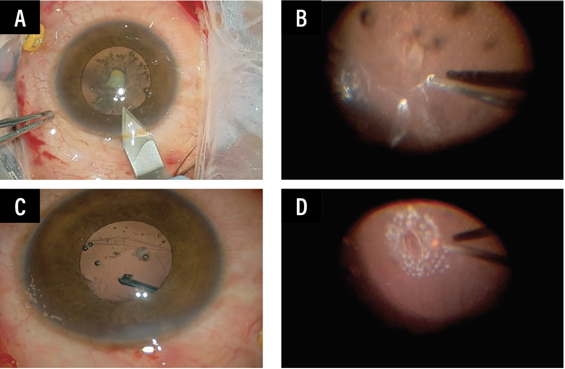 |
| Figure 1. Main tunnel for cataract extraction, IOFB removal and IOL insertion (A). The IOFB impaction site is identified and pars plana vitrectomy is completed (B). The IOFB is then brought to the anterior chamber and removed with intraocular magnet (C). Barricade laser retinopexy is done around the impaction site (D). |
Intraoperative Strategies
1. Secure the infusion port: Ensuring a stable infusion system is critical for maintaining IOP throughout the procedure.
2. Main tunnel creation: Establish a primary tunnel for cataract removal, IOFB removal and IOL placement. Creating this entry early provides a reliable route for foreign body extraction.
3. Early cataract removal: If a cataract is present, it should be removed early in the procedure to enhance visualization. Care must be taken to keep the sulcus intact for subsequent IOL placement.
4. Vitrectomy and PVD induction: Perform a thorough vitrectomy with posterior vitreous detachment induction, assisted by triamcinolone acetonide, to clear the vitreous and improve access.
5. Identifying the IOFB impaction site: Locating the exact site of IOFB impact is crucial; and the IOFB may be found near the site of impaction.
6. Ample vitrectomy around the impaction site: Carefully complete the vitrectomy around any retinal break due to IOFB impaction to reduce traction and prevent further retinal damage.
7. Locating and isolating the IOFB: Conduct a meticulous search for the IOFB, typically near the impaction site, and perform a complete vitrectomy around it to ensure safe retrieval (Figure 1).
8. Retrieving the IOFB: Using intraocular forceps (or a magnet if appropriate), gently grasp and guide the IOFB into the anterior chamber for controlled removal.
9. Exteriorizing the IOFB: Carefully extract the IOFB through the main tunnel using an intraocular magnet, maintaining control to avoid additional ocular trauma.
10. IOL placement: Once the foreign body is removed, place an IOL in the sulcus.
11. Wound closure: Securely close the main tunnel with sutures to maintain ocular integrity.
12. Endolaser barricade: Adequate laser around the impaction site to fortify the retina is paramount.
Administer antibiotics and steroids postoperatively to manage inflammation and infection risk, and follow up with imaging to monitor for complications such as retinal detachment.
This structured approach and adherence to these intraoperative strategies help to maximize safety and outcomes in IOFB cases. RS