
 | 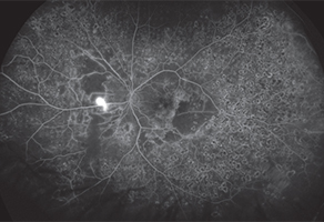 | |
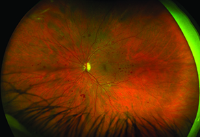
| Examples of color Optomap image (left) and fluorescein angiography (right) of diabetic retinopathy from the Optos California, which fits on a tabletop | ||
Then he saw the images. “They were actually very good,” says Dr. Brown, whose Retina Consultants of Houston has 14 offices in southeastern Texas.
The newest offering in Optos’ line of imaging systems is capable of capturing images in eight modalities, including fundus photography, autofluorescence and fluorescein angiography, along with ICG angiography. The California is a step up from the Optos 200TX, which offers all the same imaging functions except for ICG angiography.
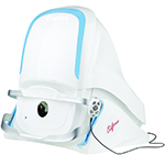
Other features of the California that appeal to Dr. Brown are its small footprint and ease of access for patients. Its compact size—the California fits on a tabletop—is a significant improvement over the original Optos platforms that required a space half the size of an exam lane. “If you had a different fluorescein camera, the California will fit in the same space if not smaller,” Dr. Brown says. “They also did some better ergonomics so it’s easier to get patients in and out.”
The device also delivers high-quality imaging out to 200° of the retinal periphery, Dr. Brown says. Changes in the software and placement of image-capturing lasers from the Optos 200TX model translate into better imaging in the superior and anterior areas. “The de-warping mechanisms also makes images more spherical as opposed to more of a warped image with other systems,” he says.
Optos California also uses browser-based software for viewing images. “While it’s always counterintuitive to use a new software program, it is much faster to go through images with the new software once you get the hang of it,” Dr. Brown says.
At first, Optos offered one version of California that included all eight imaging modalities, but it is introducing three different models within the California line at the American Academy of Ophthalmology: one with color photography and autofluorescence; another that adds fluorescein angiography; and a third that includes color photography, autofluorescence, fluorescein and ICG angiography.
“I think it’s imperative to have widefield angiography for practices that have a high percentage of patients with diabetic retinopathy or that see a lot of patients with retinal vascular disease,” Dr. Brown says.
While standard field views can capture most of the pathology in macular degeneration, “for global disease like diabetic retinopathy and retinal vascular disease, I would feel incredibly handicapped without having a widefield angiogram,” Dr. Brown says. RS


