 |
|
Bios Dr. Muste is on the retina service at Wills Eye Hospital in Philadelphia, and also practices at Mid-Atlantic Retina. |
A 72-year-old male was referred for bilateral panuveitis and posterior scleritis complicated by serous retinal detachments. Past ocular history was notable for bilateral phacoemulsification and intraocular lens implantation. His past medical history included hypertension, gout and gastroesophageal reflux disease. Past medical history was most remarkable for a history of bilateral panuveitis diagnosed two years prior to presentation secondary to syphilis. The patient had previously completed two weeks of intravenous penicillin G treatment at the time of initial diagnosis. Since that time, he was continually on oral prednisone (variable dosing, but as high as 60 mg per day). He was referred for a worsening recurrence of posterior scleritis—initially presenting in the left eye and developing in the right eye four weeks later—now representing a severe bilateral exacerbation.
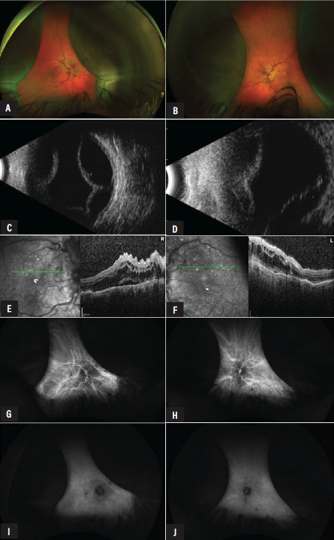 |
| Figure 1. Color fundus photos showing prominent serous choroidal detachments with shallow serous retinal detachments in the right eye (A) and left eye (B). B-scan ultrasonography confirmed both serous choroidal detachment in the right eye (C) and serous choroidal and retinal detachment in the left eye (D). Horizontal rasters on optical coherence tomography of the right eye (E) and left eye (F) revealed RPE changes, a thickened choroid, macular edema and small pockets of subretinal fluid. Early indocyanine green angiography of both eyes revealed increased tortuosity of the choroidal vessels of the right eye (G) and left eye (F) that transitioned to later diffuse hyperfluorescence in both right (H) and left (I) eyes. |
On presentation, the patient was maintained on 60 mg of prednisone daily and complained only of blurred vision and had never had ocular pain. He was counting fingers in both eyes. Intraocular pressure was 15 mmHg in the right eye and 18 mmHg in the left eye. The patient endorsed no ocular pain on his current dose of 60 mg of oral prednisone. The anterior segment examination disclosed no cells with well-positioned intraocular lenses in both eyes. The posterior segment examination was notable for a rare vitreous cell with both serous choroidal and retinal detachments in both eyes (Figure 1).
A differential diagnosis was created for a patient with serous retinal and choroidal detachments, and includes broadly inflammatory, infectious, neoplastic/infiltrative, vascular and structural causes:
• Inflammatory
• Vogt–Koyanagi–Harada (VKH)
• posterior scleritis
• sympathetic ophthalmia
• Infectious
• endogenous endophthalmitis
• syphilis
• Choroidal neoplastic/infiltrative disease
• choroidal melanoma
• choroidal metastasis
• choroidal lymphoma
• choroidal hemangioma
• paraneoplastic syndromes
• Vascular
• severe hypertensive retinopathy and choroidopathy
• preeclampsia/eclampsia
• bullous central serous chorioretinopathy
• Structural
• hypotony
• drug-associated ciliochoroidal effusion
• trauma
• glaucoma surgery or intraocular surgery
• laser photocoagulation
• Medication
• sulfa drugs
• mefenameic acid
• phendimetrazine tartrate
• buproprion
• ephedrine
• topiramate
B-scan ultrasonography confirmed serous choroidal detachments, inferior serous retinal detachments, and didn’t show evidence of any intraocular mass lesions, T-sign or retrobulbar hypoechoic lesions. A-scan ultrasonography revealed an axial length of 22.31 mm in the right eye and 22.54 mm in the left eye. Fluorescein angiography was deferred due to previously documented fluorescein allergy. Indocyanine green angiography showed evidence of early choroidal vascular tortuosity and congestion with diffuse late hyperfluorescence without evidence of a choroidal vascular lesion, hot spots or hypofluorescent choroidal lesions. Prior laboratory work-up from the referring doctor was reviewed and showed a positive FTA-Abs, with a low RPR titer of 1:2. His RPR titer was repeated following intravenous penicillin therapy and remained stable at 1:2. Additional testing from the referring doctor disclosed the following:
• negative Lyme antibodies;
• HIV testing;
• QuantiFERON gold;
• angiotensin-converting enzyme;
• rheumatoid factor;
• C-reactive protein;
• erythrocyte sedimentation rate;
• human leukocyte antigen-B27;
• antinuclear antibody;
• antineutrophil cytoplasmic antibody testing
• an unremarkable complete metabolic panel;
• complete blood counts; and
• chest X-Ray.
Blood pressure was checked in office and the patient was normotensive. His medication list was thoroughly reviewed, and no offending agents were identified.
Given the patient’s chronic presentation without ocular pain and the absence of evidence of intraocular inflammation or infection, intraocular neoplasm, hypotony or a drug-associated adverse effect, as well as well-controlled hypertension and appropriately treated syphilis, a tentative diagnosis of uveal effusion syndrome was made by exclusion. However, his serous choroidal and retinal detachments persisted on high-dose steroids.
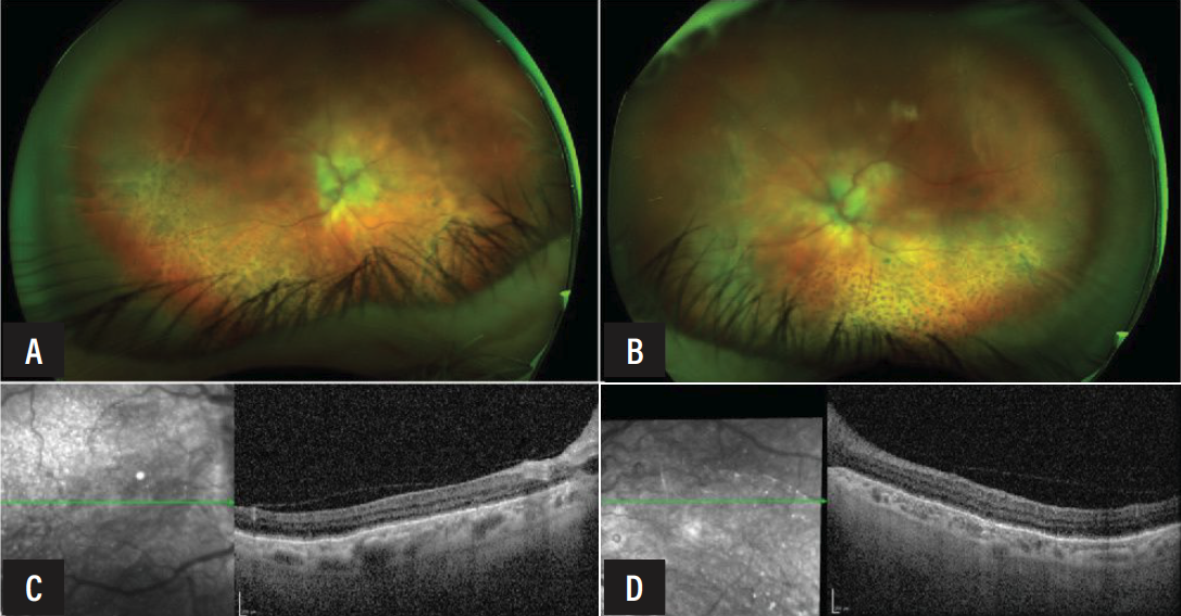 |
| Figure 2. Color fundus photograph of the right eye (A) and left eye (B) demonstrating resolution of the serous retinal and choroidal detachments with subsequent demarcation lines and diffuse pigment mottling. Optical coherence tomography of the right eye (C) and left eye (D) revealing resolution of the subretinal fluid and macular edema. |
The decision was made to initiate a taper of oral steroids by 10 mg a week and proceed with four scleral windows with sclerostomies in the right eye, and, if successful, proceed in the left eye (See surgical video at https://bit.ly/4ySzqg8). Intraoperatively, his sclera was noted to be abnormally thickened and spongiform OU. His postoperative course was unremarkable, with the choroidal detachment resolving at one week after surgery and the serous detachments resolving at one month in both eyes. At his final postoperative visit nearly 11 months after scleral window, the vision improved to 20/100 in the right eye and remained stable at CF in the left. His examination showed demarcation lines and diffuse pigment mottling; however, no serous retinal detachment was appreciated (See Figure 2).
Discussion
Uveal effusion syndrome is a rare, frequently bilateral disorder best understood as the abnormal accumulation of protein-rich fluid within the suprachoroidal and supraciliary spaces, which generates serous detachment of the choroid and ciliary body, and subsequently the retina. Because choroidal melanoma, metastasis, posterior scleritis, VKH disease, and central serous chorioretinopathy can each reproduce elements of the picture, UES is fundamentally a diagnosis of exclusion.1,2 This article reviews the pathophysiology, presentation, diagnosis and treatment of UES.
Pathophysiology
In brief, UES can be summarized as an abnormal collection of fluid that expands the suprachoroidal space, elevating the choroid and ciliary body and secondarily detaching the overlying neurosensory retina.3-5 The histopathology of resected sclera in affected eyes shows disorganized collagen fiber bundles and deposition of glycosaminoglycans and proteoglycans within the extracellular matrix.2,4 This abnormal, thickened sclera resists the normal transscleral
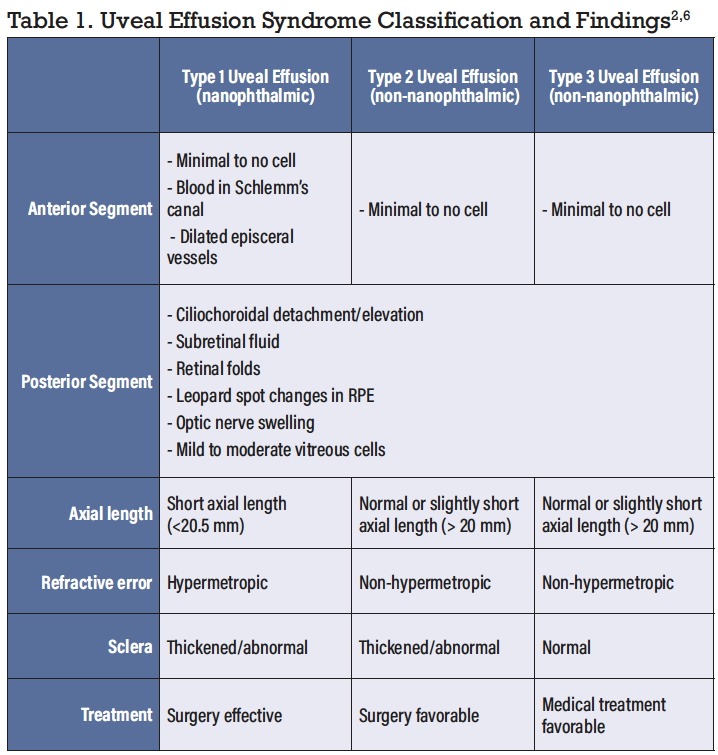
egress of albumin and other macromolecules from the suprachoroidal space. These retained oncotically active proteins then attract and retain fluid. Accordingly, Osaka, Japan’s Masanobu Uyama, MD, proposed a framework for better understanding causes of UES (See Table 1).
As discussed below, the distinction has a role in treatment. Types 1 and 2 share the scleral pathology that scleral surgery is designed to address. Type 3 eyes may be better treated by initial medical therapy.2,4,6
Presentation
UES classically affects otherwise healthy middle-aged, hypermetropic men.1 One eye may become symptomatic months to years before the fellow eye follows. Involvement is bilateral in roughly two-thirds of patients. Patients present with painless (89 percent) blurring or metamorphopsia as subretinal fluid tracks into the macula.7 Patients may give a history of relapsing-remitting vision loss with spontaneous exacerbations and partial remissions that can span years.
Examination discloses bullous, non-rhegmatogenous retinal detachment with characteristic shifting fluid. It’s possible that annular ciliochoroidal detachment and dilated episcleral vessels may also be present. Though a small number of vitreous cells may be present, there’s no robust inflammatory reaction in the posterior or anterior chamber. Intraocular pressure is typically normal. However, in the nanophthalmic eye, a short axial length and crowded anterior segment predispose to secondary angle closure; in these patients, acute or chronic angle-closure glaucoma may precede recognition of the effusion.7,8
Diagnostic features
UES remains a diagnosis of exclusion. The first task is to rule out the conditions it imitates: primary choroidal neoplasms and metastasis; posterior scleritis; VKH disease; choroiditis; CSCR; hypotony; drug related adverse effects; hypertensive retinopathy; and choroidopathy. A careful history, ocular examination, lab work, and multimodal imaging can aid to that end.
As part of a complete ocular examination, emphasis should be placed on searching for cell and retinal breaks that would suggest other etiologies. Over time, chronic subretinal fluid produces the characteristic “leopard-spot” pattern of the retinal pigment epithelium: scattered, mottled yellow-orange deposits that are a secondary consequence of long-standing detachment rather than an acute sign. Because they develop only after the fluid has been present for some months, their absence early doesn’t exclude the diagnosis, and their persistence after resolution shouldn’t be mistaken for active disease.6,7
 |
Multimodal imaging both supports the diagnosis and helps to exclude mimics. B-scan ultrasonography demonstrates the combination of choroidal and retinal detachment, frequently as an extended, multilayered and kinetically mobile detachment, and can show scleral thickening.9 Simultaneously B-scan excludes a choroidal mass and identifies the “T-sign” that signals posterior scleritis. Additionally, an A-scan supplies the accurate axial length needed to identify nanophthalmos. Ultrasound biomicroscopy can reveal an annular peripheral ciliochoroidal detachment and a characteristically spongy, edematous ciliary body, sometimes with interstitial lacunae.9
Fundus autofluorescence will show mixed hyper- and hypoautofluorescent pattern. Fluorescein angiography highlights the “leopard spots” as a mottled pattern of hyper- and hypofluorescence corresponding to the RPE changes. Crucially, it’ll confirm the absence of pinpoint or expansile leakage that defines CSCR.6,7 Indocyanine green angiography shows diffuse granular choroidal hyperfluorescence, most evident in the early phase, in keeping with a generalized choroidal process rather than a focal leak. Optical coherence tomography can appreciate subretinal fluid, RPE undulations or folds and increased choroidal thickness, and is useful for tracking the macula through treatment.
Auxiliary lab work can help ensure there’s no underlying infectious disease with protean manifestations, particularly tuberculosis or syphilis. A CT or MRI can provide a further check against an occult mass. In certain instances, MRI (T1) with gadolinium can show uveal detachment.7 Though less generalizable, clinicians may consider genetic testing in the nanophthalmic (Type 1) subtype. Five genes (i.e., MFRP, TMEM98, PRSS56, BEST1 and CRB1) and two loci have been implicated in familial forms of nanophthalmos.10 Knowledge of this inheritance pattern can facilitate genetic counseling for familial forms of this condition and may help to decrease amblyopia from uncorrected hyperopia, heighten monitoring and prevent vision loss from complications.
Treatment
Management is individualized and guided by Uyama subtype, disease severity and symptomatic impact. As illustrated in the case presented here, Type 2 eyes benefit from quadrantic partial-thickness scleral windows placed anterior to the equator.4,5 Type 1 eyes also benefit from this intervention. Vortex vein decompression is an alternative but carries a higher risk of vein amputation and hemorrhage, and most surgeons favor sclerectomy.3-5,11 The anatomic results in these subtypes are good. Recent series and a systematic review confirm high reattachment rates with quadrantic lamellar sclerectomy and sclerostomy, alongside meaningful visual recovery.12
Type 3 eyes may be better approached with an initial medical therapy. In the largest reported experience, Wills Eye Hospital’s Carol Shields, MD, and colleagues managed 104 eyes with corticosteroids delivered by oral, periocular or topical routes, or with observation; using these approaches, disease control was achieved in the large majority, and scleral window surgery was required in only about 5 percent, with some eyes resolving spontaneously.7
Additional considerations for management include high risk of angle-closure glaucoma from anterior-segment crowding that may require referral or management by cataract extraction or laser peripheral iridotomy. Finally, given the relapsing course and frequent bilaterality, long-term follow-up is essential.
Conclusion
Uveal effusion syndrome is a diagnosis of exclusion best evaluated using a variety of tools at the retina specialist’s disposal. B-scan ultrasonography aids diagnosis and, together with angiography, helps distinguish UES from possible mimics. Eyes with nanophthalmos or abnormal sclera (types 1 and 2, respectively) are generally best approached with scleral surgery, while eyes with a normal sclera (type 3) more often respond to corticosteroids. RS
REFERENCES
1. Sharma R, Foot B, Jackson TL. A prospective, population-based, surveillance (BOSU) study of uveal effusion syndrome in the UK. Eur J Ophthalmol 2021;31:5:2451-2456.
2. Uyama M, Takahashi K, Kozaki J, et al. Uveal effusion syndrome: Clinical features, surgical treatment, histologic examination of the sclera, and pathophysiology. Ophthalmology 2000;107:3:441-449.
3. Schepens CL, Brockhurst RJ. Uveal effusion. 1. Clinical picture. Arch Ophthalmol 1963;70:189-201.
4. Gass JD. Uveal effusion syndrome: A new hypothesis concerning pathogenesis and technique of surgical treatment. Trans Am Ophthalmol Soc 1983;81:246-260.
5. Johnson MW, Gass JD. Surgical management of the idiopathic uveal effusion syndrome. Ophthalmology 1990;97:6:778-785.
6. Li HH, Hunter KC, Thomson AC, Hunter AA. Medical therapy and scleral windows for uveal effusion syndrome: A case series and literature review. Ophthalmol Ther 2023;12:
1)35-53.
7. Shields CL, Roelofs K, Di Nicola M, Sioufi K, Mashayekhi A, Shields JA. Uveal effusion syndrome in 104 eyes: Response to corticosteroids – The 2017 Axel C. Hansen lecture. Indian J Ophthalmol 2017;65:11:1093-1104.
8. Elagouz M, Stanescu-Segall D, Jackson TL. Uveal effusion syndrome. Surv Ophthalmol 2010;55:2:134-145.
9. Fledelius HC, Fuchs HJ, Jensen PK, Scherfig E. Uveal effusion and ultrasonic imaging: A clinical series. Acta Ophthalmol Scand 2002;80:2:202-210.
10. Carricondo PC, Andrade T, Prasov L, Ayres BM, Moroi SE. Nanophthalmos: A review of the clinical spectrum and genetics. J Ophthalmol 2018;2018:2735465.
11. Brockhurst RJ. Nanophthalmos with uveal effusion: a new clinical entity. Trans Am Ophthalmol Soc 1974;72:371-403.
12. Braga de Sousa L, Barbosa-Breda J. Sclerectomies in nanophthalmos and idiopathic uveal effusion syndrome: A systematic review. Graefes Arch Clin Exp Ophthalmol Albrecht Von Graefes Arch Klin Exp Ophthalmol 2025;263:10:2709-2722.